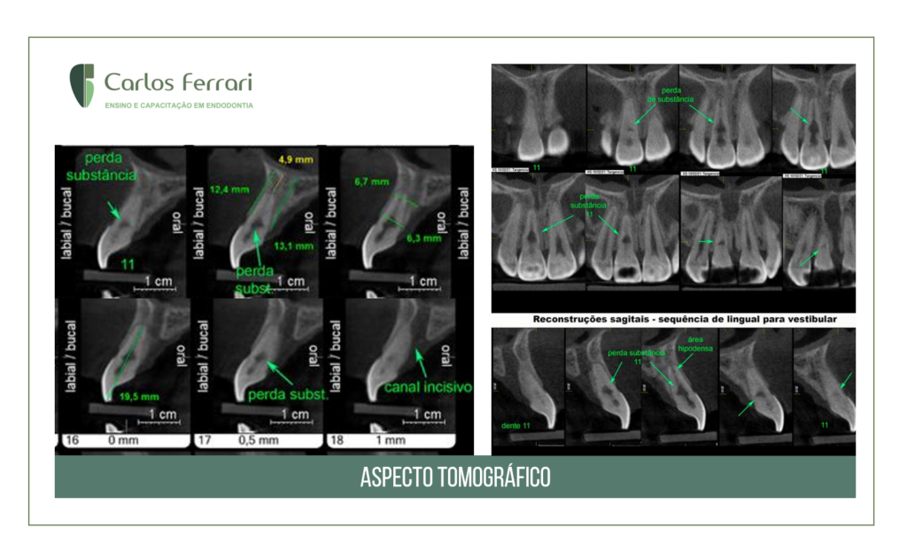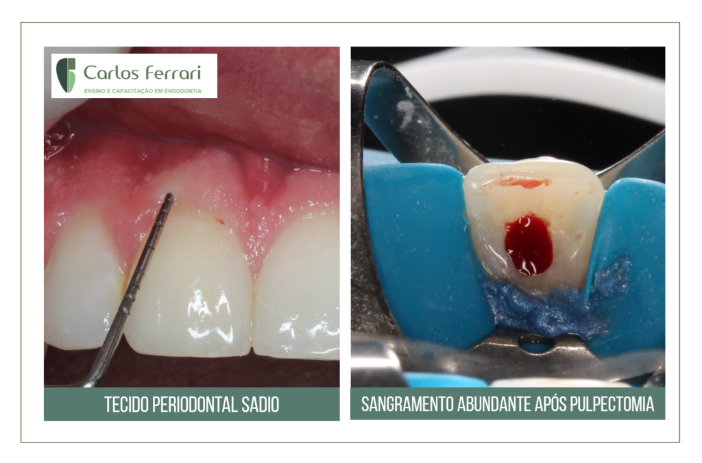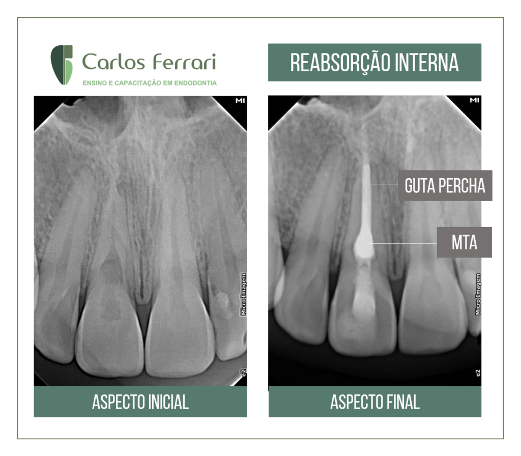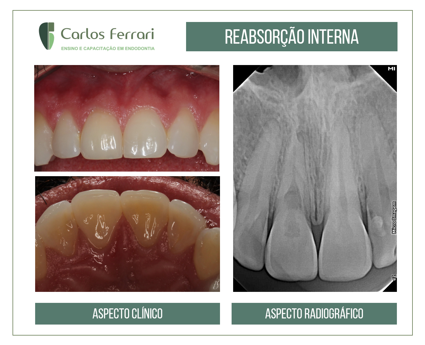
Male patient, 30 years old, with a history of dental trauma. On clinical examination, thermal tests, percussion and palpation were normal on all upper anterior teeth, and there was no periodontal pocket or evidence of gingival inflammation. Radiographic examination revealed an image suggestive of internal inflammatory root resorption. A CT scan was then ordered to verify the extent of the lesion and possible periodontal origin, for differential diagnosis of external cervical resorption. Treatment was performed with gutta-percha obturation in the apical 8 mm of the canal and the resorption filled with MTA (Angelus).
Patient seen in the specialization clinic of class I in endodontics of the Hodos School in Brasília, by students Jéssica and Erika.