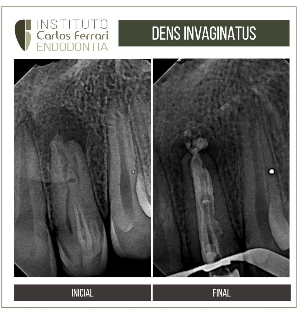
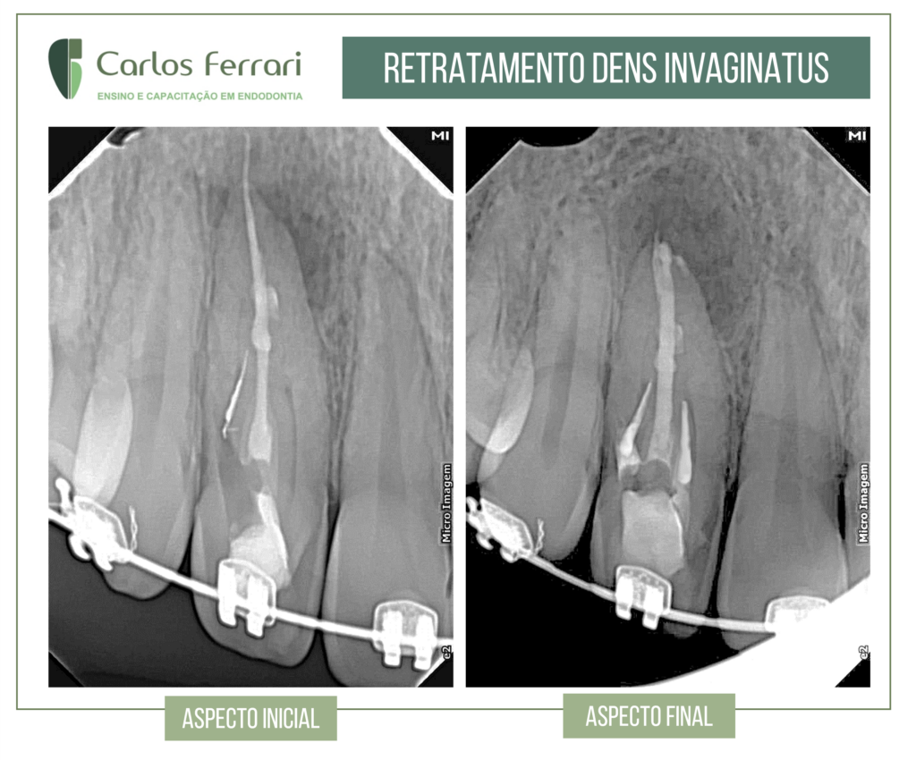
Endodontic treatment of dens invaginatus
Endodontic treatment of dens invaginatus.
Endodontic treatment of dens invaginatus Patient came to the clinic complaining of swelling and severe pain on the left side of the face. After...


Endodontic treatment of dens invaginatus Patient came to the clinic complaining of swelling and severe pain on the left side of the face. After...
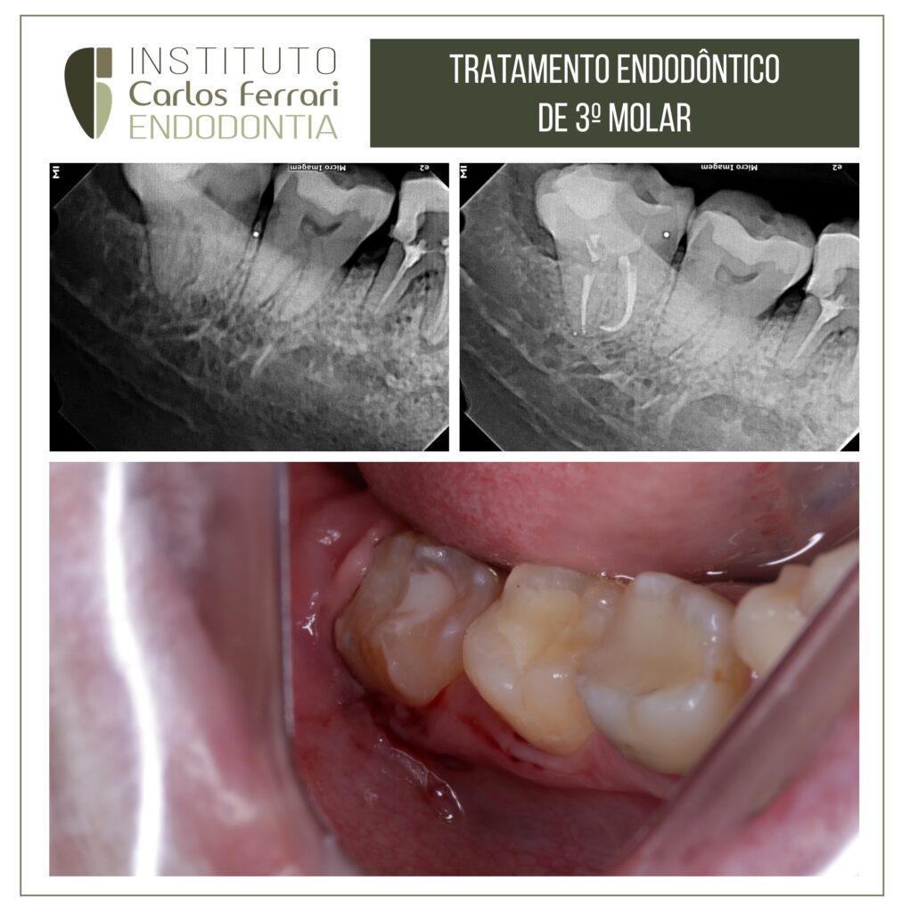
Third molar endodontic treatment. Endodontic treatment of third molars in a full arch is rare, especially in male and female patients....
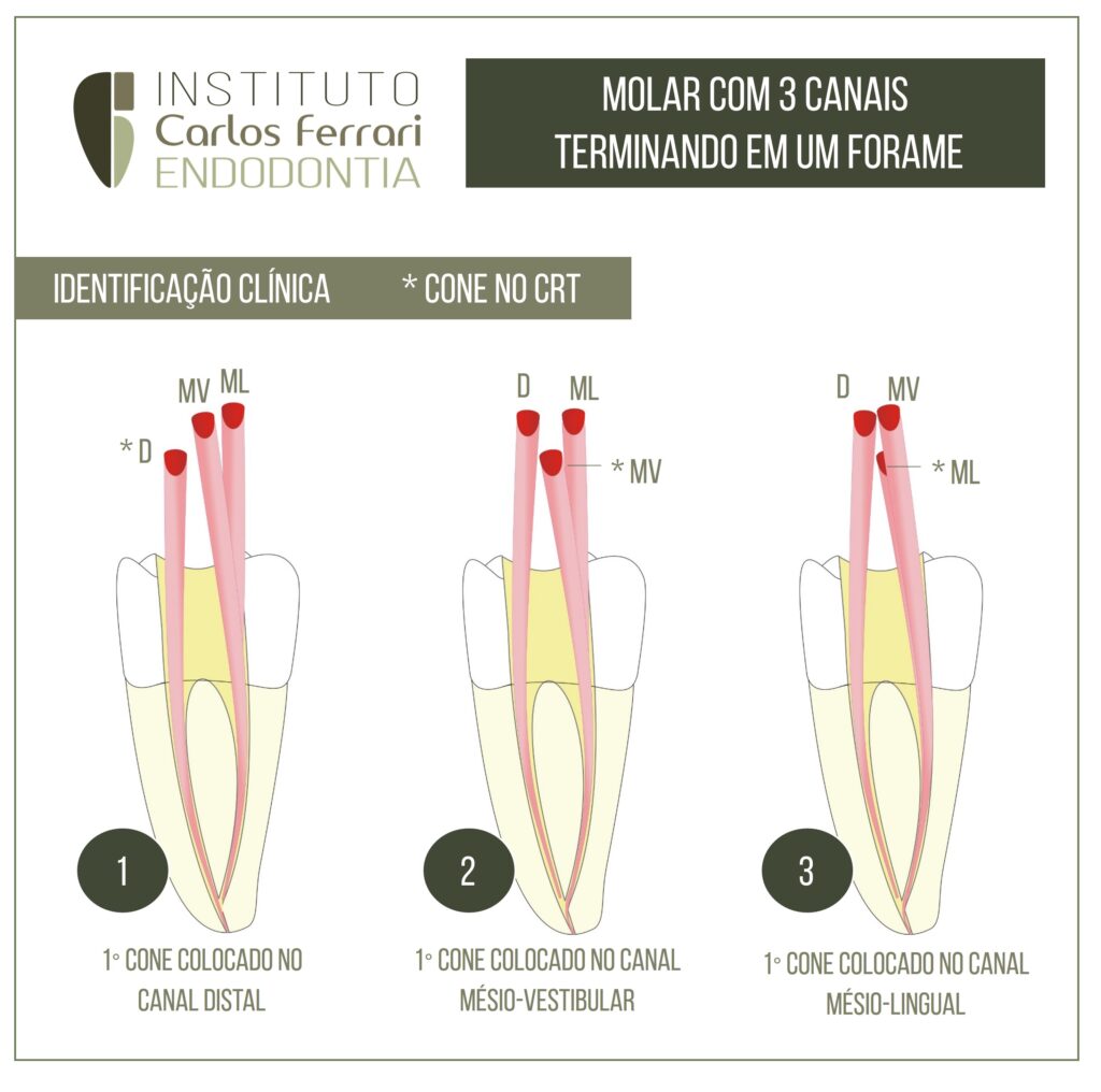
Anatomy in endodontics. Molar with 3 canals ending in a foramen. Many times, simple clinical maneuvers allow us to solve doubts that...
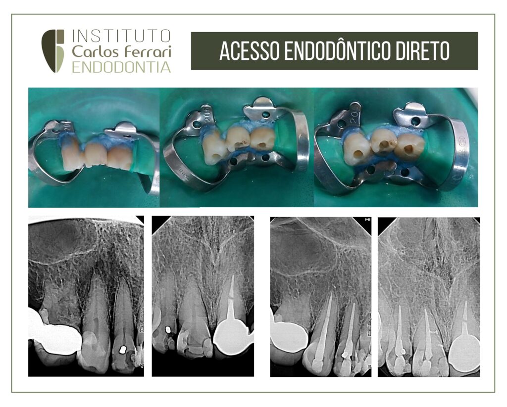
Access surgery. Patient indicated for endodontic treatment of the upper anterior teeth for aesthetic prosthetic rehabilitation. In this way it was considered...
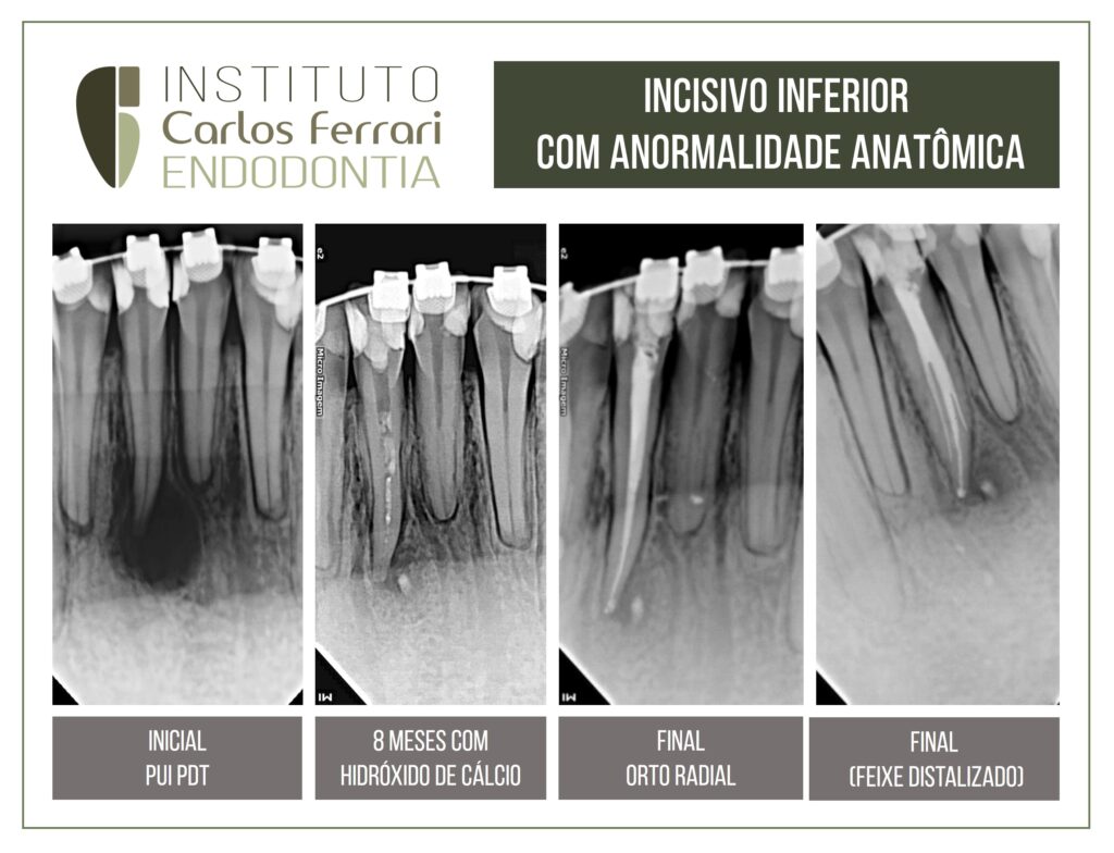
Dental anatomy. Anatomical abnormality of the lower lateral incisor tooth. Patient sought care for endodontic treatment of tooth 41 indicated...
Treatment of dens invaginatus with MTA. Patient came to the clinic complaining of pain on palpation in the right upper gingiva and...
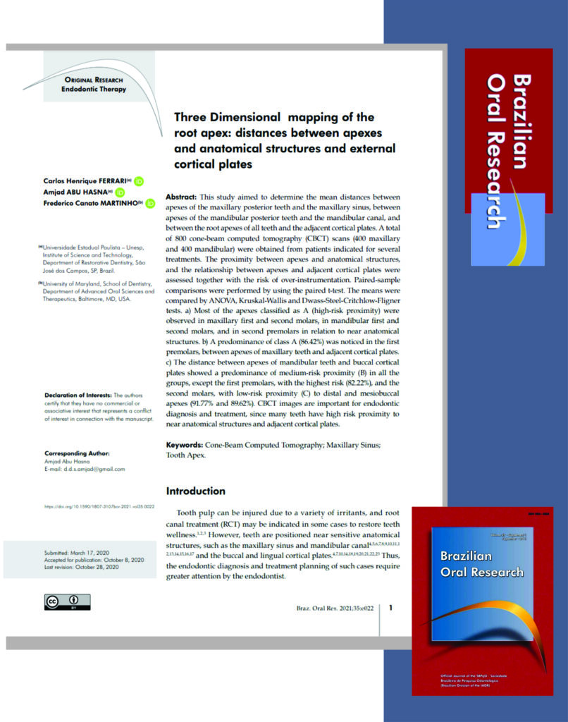
Paper published in the Brazilian Oral Research Journal, with colleagues Amjad Abu Hasna and Frederico Canato Marinho. Related topics: cone beam tomography in endodontics.
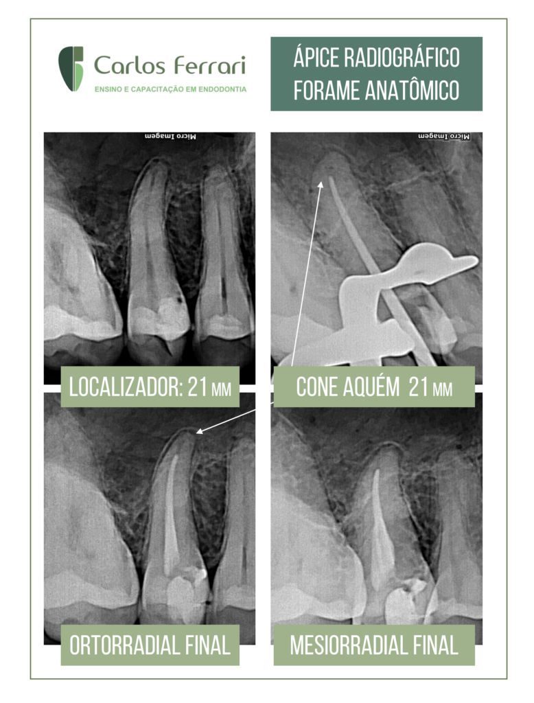
Small doubts from the clinical routine. The locator points 21mm and on the cone probe, also adjusted for this length, it seems well short of the ideal. Odontometry is repeated and the same result is obtained. When obturating, the final radiograph also seems unsatisfactory, but in mesial view we can get a better idea of the apical positioning of the cone. This is why it is important to rely on a good apical locator and to know the anatomy in the region of the foramen. Most of the time it does not coincide with the radiographic vertex.
Endodontic anatomy of the lower canine. Patient came to the clinic with a complaint of mild pain when biting on the region of the...