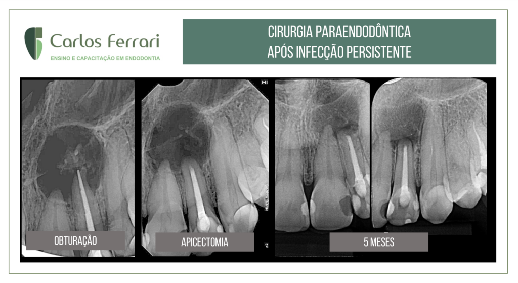
Paraendodontic surgery after persistent infection in a tooth with periapical lesion.
Despite all the modern resources in endodontic treatment, in some cases the conventional treatment is not enough...


Despite all the modern resources in endodontic treatment, in some cases the conventional treatment is not enough...
The update course in endodontics in Taubaté was a success. We thank Chibebe Cursos and the students for the moments of pleasure and...

With honor, I will participate tomorrow in the evaluation of the papers of the XXXIII Scientific Initiation Congress of UNESP SJC. Thanks to Prof. ....
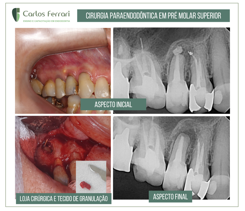
Paraendodontic surgery of periapical lesion in upper second premolar. Patient referred by a specialist colleague after non-remission of the fistula...
There are cases when we need to analyze the treatment alternatives taking into account several factors. This patient was referred by...
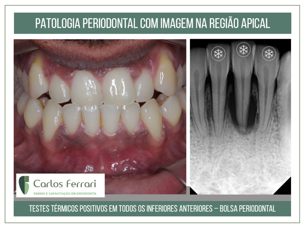
Patient sent for endodontic treatment of element 31 due to observation of periapical radiolucent image. On clinical examination, the tests revealed no different response in relation to adjacent teeth, positive in relation to the thermal test with gas and negative to percussion. Slight pain was reported in relation to the palpation test in the root region of tooth 31. On probing, periodontal pocket was revealed in the proximal regions. The presence of calculus was also observed. The pathology was diagnosed as of periodontal origin and the case was sent to the periodontist for treatment and orientation for possible endodontic treatment concomitant with periodontal.

Learn and update yourself on the latest literature on irrigation during endodontic treatment. Irrigating substances, techniques, protocols, and accident prevention.Topics:IntroductionIrrigating substancesIrrigant interactionIrrigation techniquesIrrigation protocolProf. Dr. Carlos Ferrari PhD in EndodonticsSpecialist in EndodonticsSpecialist in RadiologySpecialist in Orofacial Pain
Theoretical laboratory course for dental students. Emphasis on undergraduate endodontics with introduction to automated preparation. Anatomy and access. Manual and automated preparation. Irrigation. Obturation. Diagnosis and urgency.
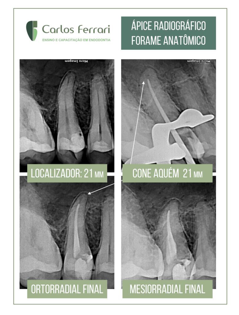
Small doubts from the clinical routine. The locator points 21mm and on the cone probe, also adjusted for this length, it seems well short of the ideal. Odontometry is repeated and the same result is obtained. When obturating, the final radiograph also seems unsatisfactory, but in mesial view we can get a better idea of the apical positioning of the cone. This is why it is important to rely on a good apical locator and to know the anatomy in the region of the foramen. Most of the time it does not coincide with the radiographic vertex.
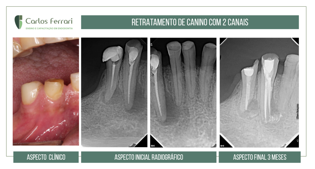
Endodontic anatomy of the lower canine. Patient came to the clinic with a complaint of mild pain when biting on the region of the...