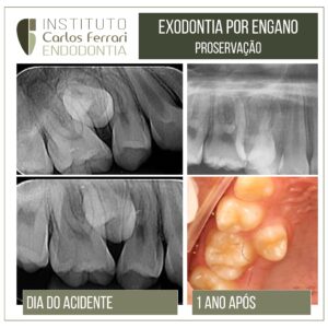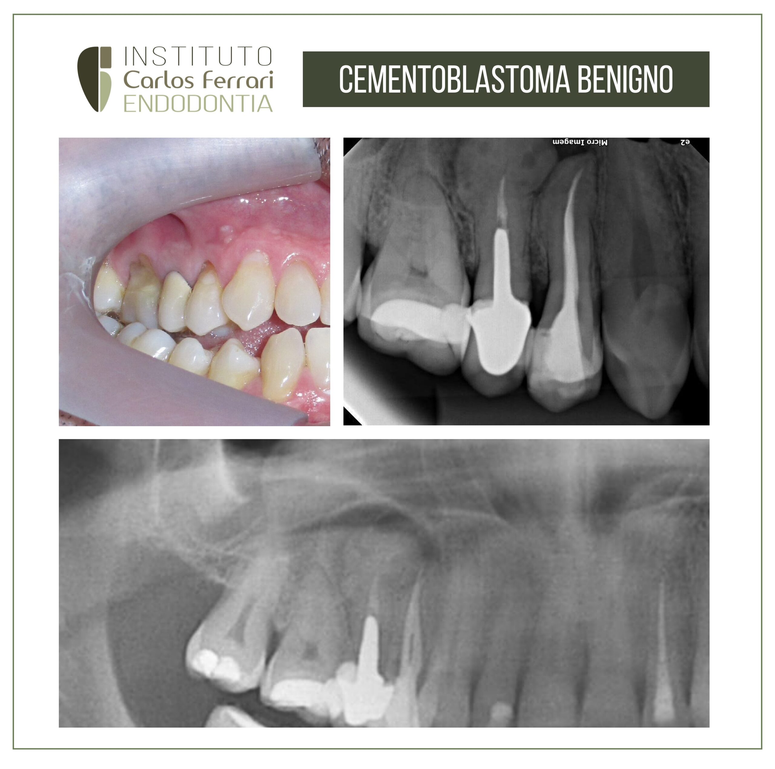
Benign cementoblastoma. Case report.
Caucasian patient, male, 35 years old, was referred by a colleague for evaluation and possible endodontic surgery in tooth 15, with endodontic treatment performed and intraradicular retainer installed. On clinical examination, a well-adapted prosthesis on the tooth and the presence of scar tissue in the periapical region of tooth 14, possibly related to a fistula originated from an endodontic infection of this tooth, recently treated.
On periapical and panoramic radiographic examination of the region, a circumscribed circular radiopaque image of about 15 mm in diameter is observed, with small radiolucent areas inside and limited by a radiolucent halo of about 2 mm throughout its apical and middle extent, and with its cervical edges with the image merging with the image of the root of tooth 15.
The initial diagnostic hypothesis was benign cementoblastoma, a lesion of non-endodontic origin, whose most recommended treatment is the removal of the element and the lesion, especially due to its large cervical extension. The patient was referred to a stomatologist, who removed the specimen and confirmed the diagnosis on pathological examination.
The case alerts to the attention to non-endodontic lesions, which are often treated as such for lack of a refined diagnosis or ignorance of the pathologies that have images in the periapical region.
Benign cemento blastoma. In: Neves et al. ev. cir. traumatol. buco-maxilo-fac. vol.10 no.2 Camaragibe Abr./Jun. 2010:
INTRODUCTION
Benign Cementoblastoma (CB) is a pathology derived from neoplastic cementoblasts, originating from the periodontal ligament1, and its etiology is still unknown, as are those of other odontogenic tumors.
According to the classification of the World Health Organization (2005), CB along with Odontogenic Myxoma and Odontogenic Fibroma are neoplasms formed by odontogenic ectomesenchyme, with or without odontogenic epithelium. It is a relatively rare entity, with just over a hundred reported cases, comprising 0.8% to 2.6% of odontogenic tumors.
CB presents as a mass, formed by cementum-like material, adhering to the tooth root and causing root resorption, while maintaining the pulp vitality of the unit involved.
Most of the data on this pathology has been extracted from case reports and the publication of small series, so information about the lesion is still limited, which makes any additional information of utmost importance in order to outline the profile of the condition.
The objective of this article is to report a case of CB, diagnosed in a routine radiographic examination, discussing relevant aspects about its clinical and radiographic characteristics, and also its treatment.
Benign Cementoblastoma