

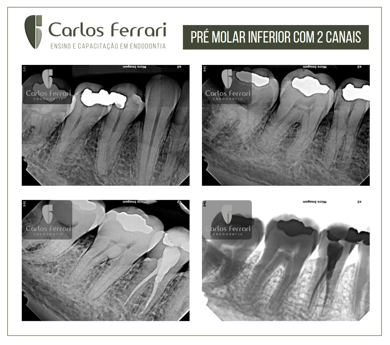
Mandibular premolar. Endodontic treatment of tooth 45 with two canals
in: https://www.forp.usp.br/restauradora/Anat.htm
The anatomy of the root canal system dictates the parameters under which endodontic treatment will be performed and affects the chances of success. This anatomy of each tooth has common characteristics as well as very complex variations.
The tooth radiograph can reveal much of the internal anatomy that, associated with theoretical knowledge, dictates the size of the drill to be used in the access surgery, its direction, the size of the first instrument to be used inside the root canal, and also which modifications should be used to prepare the endodontic cavity in order to facilitate the location of the root canals.
Thus, the knowledge of the anatomy of the root canals helps the professional a lot, from the access surgery to the obturation of the canals and it is a safe route to obtain a lot of success and avoid unpleasant situations. In this text we will recall, in a very simple way, the internal anatomy of each group of teeth.
In this article, we will give special focus to the developmental anomalies that can occur in human teeth.
Lower Premolars
First Premolar Inferior
The mandibular first premolars are probably the most difficult to treat endodontically because they have a very complex internal and external anatomy.
They can present themselves as follows: (a) one canal and one foramen; (b) one canal that bifurcates in the apical third, ending in two independent foramens; (c) one canal that bifurcates in the middle third of the root ending in two independent foramens; (d) two separate canals from the cervical third of the pulp cavity , ending in two independent foramens, (e) two separate canals from the cervical third, ending in a single foramen; (f) two canals that bifurcate in any of the thirds and end in a single foramen. Besides these anatomical variations, they can also have fused roots with two, three or four canals.
Once again, we insist on the attention that must be given to the study of the diagnostic radiographs before starting the endodontic treatment of these teeth. Often, depending on the number of canals present, we must enlarge the access cavity to the pulp chamber in the buccal-lingual direction, to facilitate instrumentation and obturation of the root canals.
Second Premolar Inferior
The anatomy of these teeth can be very complex, similar to the mandibular first premolars.
https://ferrariendodontia.com.br/tratamento-endodontico-hipercementose/
