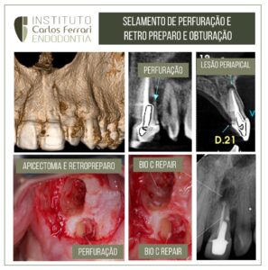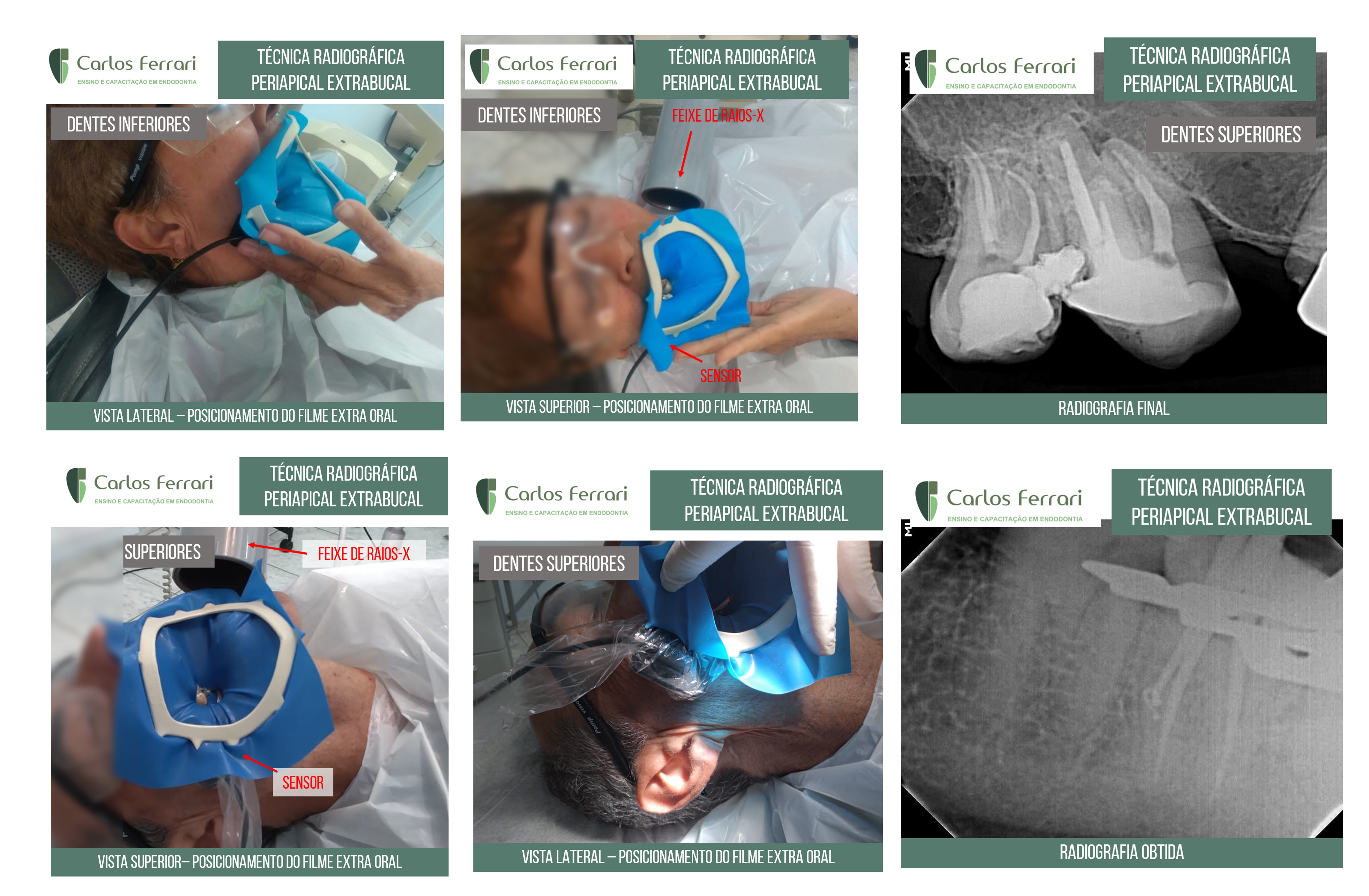
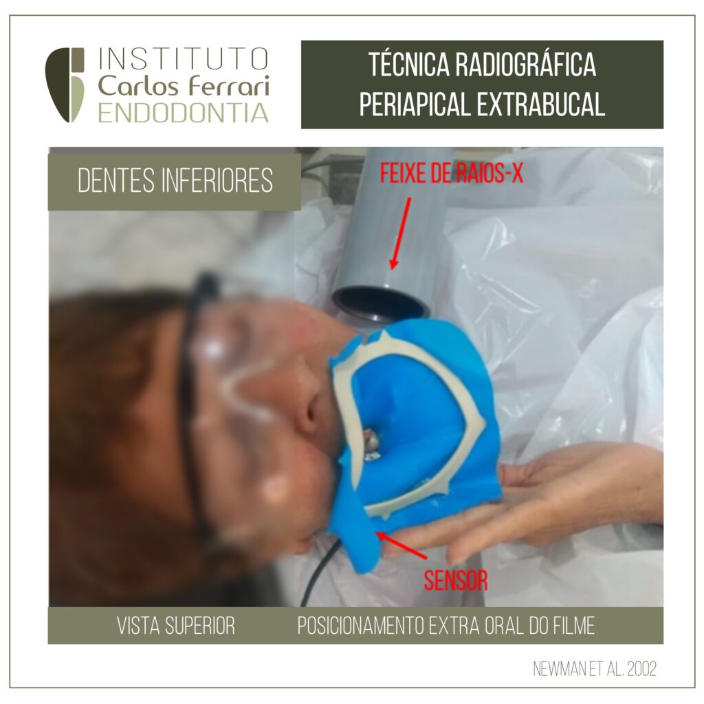
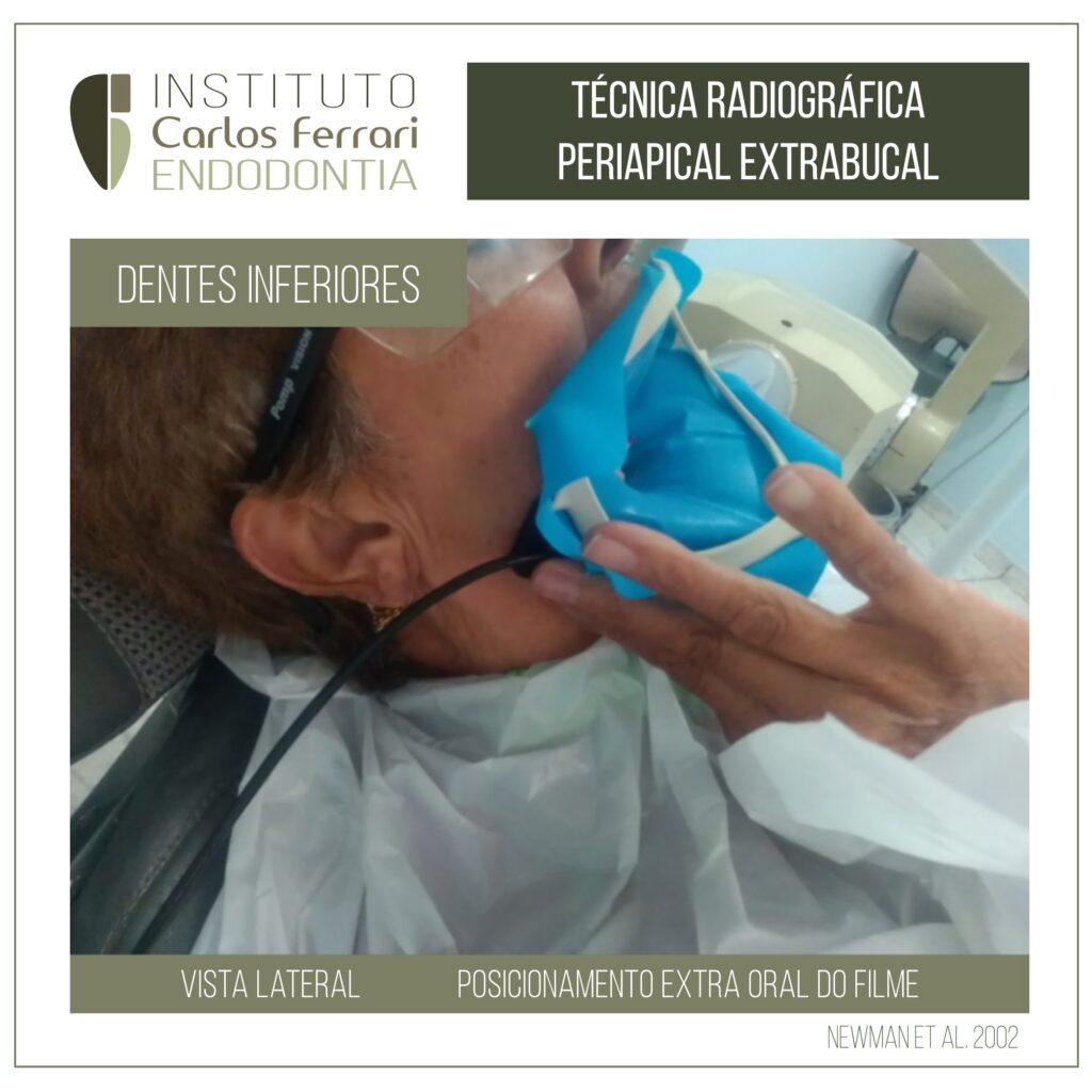
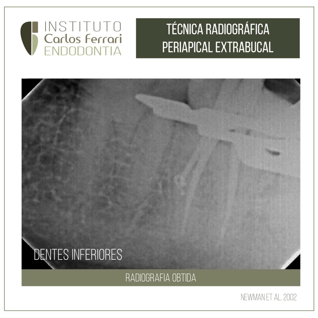
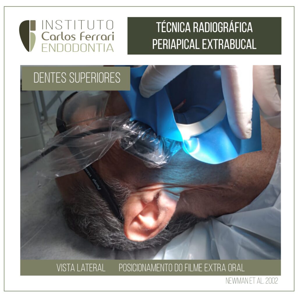
Casos em que os pacientes sentiam náuseas durante a tomada radiográfica com isolamento absoluto. Optou-se pela radiografia periapical posicionada fora da boca do paciente, de acordo com proposto por Newman et al., 2002. O resultado permite a interpretação, embora com maiores sobreposições de imagem.
Radiografia periapical extrabucal
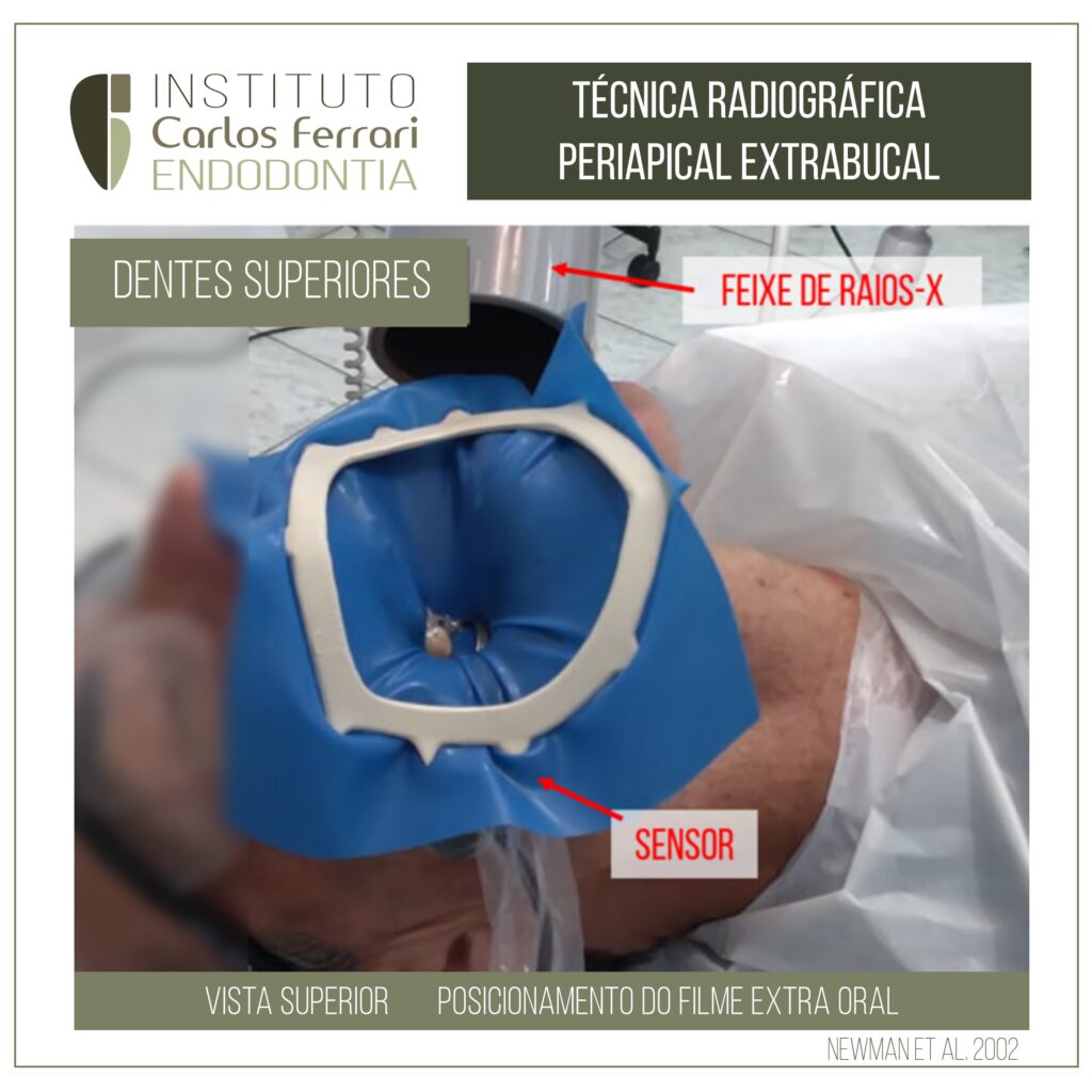

In: Newman et al. Extraoral Radiographic Technique: An Alternative Approach. Journal of Endodontics. Vol. 29, 5, Jun 2003.
Introdução: A necessidade de radiografias em todas as fases da terapia endodôntica está bem estabelecida. O clínico tem uma variedade de auxílios para facilitar uma radiografia diagnóstica. A maioria dessas ajudas (ou seja, suportes Rinn XCP , snap-a-ray, etc.) dependem de radiografia intraoral convencional. Nesta técnica, o filme é colocado lingual ao dente. O cone de raios-X é colocado diretamente vestibular ao dente, fazendo com que o feixe de raios-X atravesse os tecidos, expondo o filme (1).
Alguns pacientes não toleram oa tpecnica intraoral convencional. Esse grupo aumentou de tamanho com o advento da
radiografia digital. O sensor digital é maior e mais rígido do que um filme de raios X padrão. Os autores descobriram que certos pacientes têm dificuldade com o “volume adicionado” do sensor. Um procedimento alternativo pode ser utilizado durante a realização da terapia endodôntica para esses pacientes.
As possíveis indicações para esta técnica alternativa incluem:
- Pacientes com deficiência intelectual
- Pacientes com reflexo de vômito exagerado
- Pacientes pediátricos
- Pacientes com fobia dentária
- Pacientes com trismo