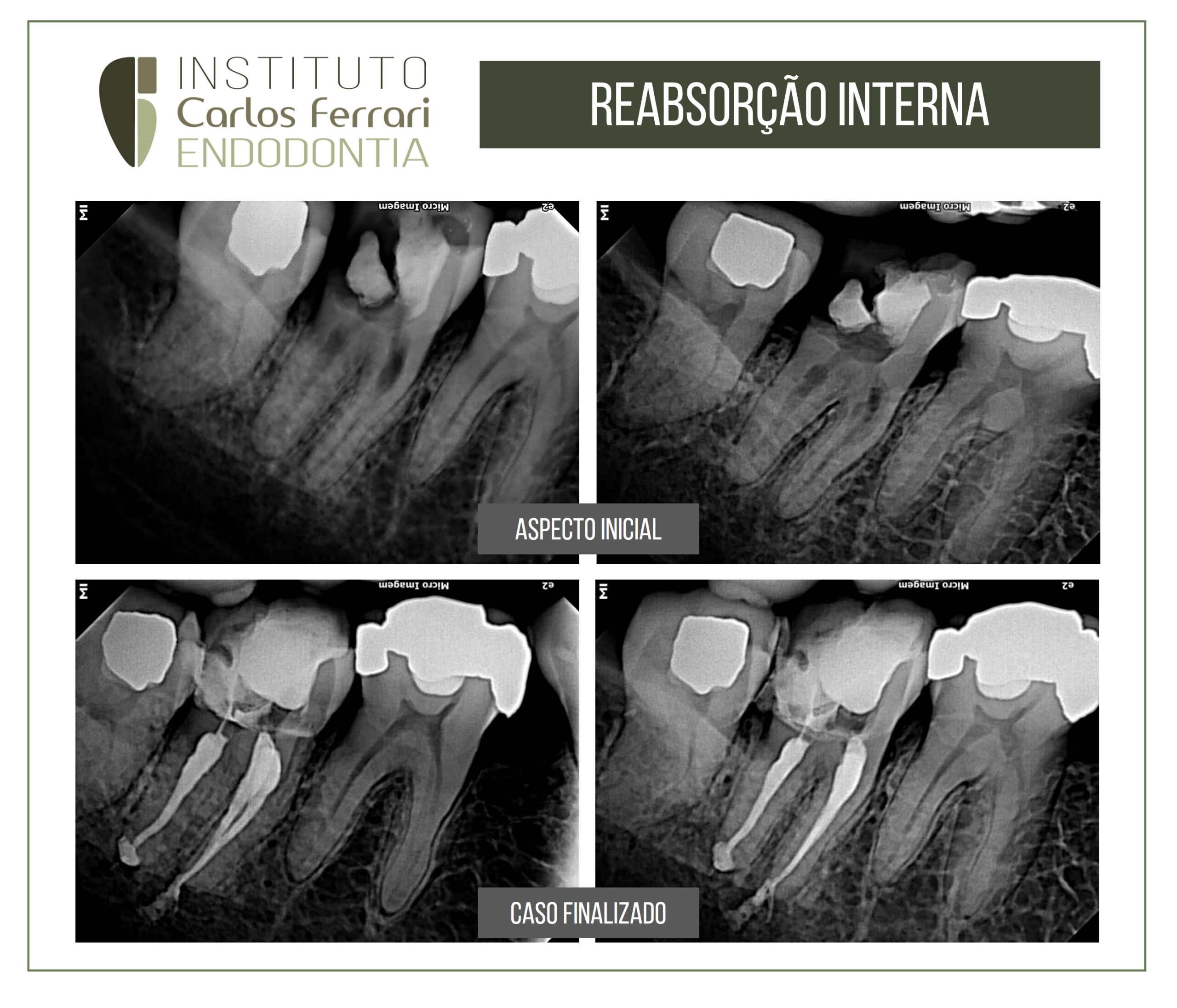
Internal root resorption in lower molar.
Patient indicated for treatment with restorative purpose.
On radiographic examination, internal resorption was noted in both roots. The obturation was performed by Tagger's technique, with a single cone, aiming to fill the gaps correctly.
Internal root resorption in: Endo, Gonçalves, Morais, Kitayama, Martinhp, Pavan, 2015. MUDI Archives, v19, n2-3, p. 43-52.
Introduction
According to the American Endodontic Association glossary, root resorption is defined as a condition associated with a physiological or pathological process that results in the loss of dentin, cementum, or bone (NE; WITHERSPOON; GUTMANN, 1999).
Physiological resorption occurs in the deciduous dentition during exfoliation and allows the eruption of its successor permanent tooth (HAROKOPAKIS-HAJISHENGALLIS, 2007; PATEL; KANAGASINGAM; PITT FORD, 2009). On the other hand, pathological resorption can occur after traumatic injuries, orthodontic movement, chronic inflammation of
infectious origin of pulp or periodontal tissues, surgical procedures, and excessive pressure from an impacted tooth or a tumor (FUSS; TSESIS; LIN, 2003).
Resorptions can be classified into internal or external resorption. Internal root resorption is an inflammatory process that begins on the inner surface of the pulp cavity with the loss of dentin, and may reach the cementum (FUSS; TSESIS; LIN, 2003). Its etiology is not fully established, and trauma is the main etiological agent CALISKAN; TURKUN, 1997). While the external root resorption is a loss of tooth structure, initiated by a mineralized or denuded area of the root surface.
Both resorptions depending on their progression can cause irreversible damage to the tooth structure, requiring adequate treatment and monitoring. Since inflammatory root resorption is usually of infectious origin, antimicrobial strategies should be applied to improve prognosis (CVEK, 1973; SJOGREN et al., 1997).
In many clinical situations, periapical radiographs do not allow a safe and accurate diagnosis of tooth resorption. However, there are cases in which the identification of the type of resorption, its degree of evolution, limits and cause cannot be definitely determined.
Thus, cone beam computed tomography is an additional resource in the detection of root resorption (COHENCA et al., 2007; REN et al., 2013), and has low radiation dose when compared to conventional computed tomography, besides including advantages such as better accuracy and resolution. This technology offers an image in three dimensions, eliminating the overlapping of images as observed in periapical radiographs(COTTON et al., 2007).
Internal root resorption