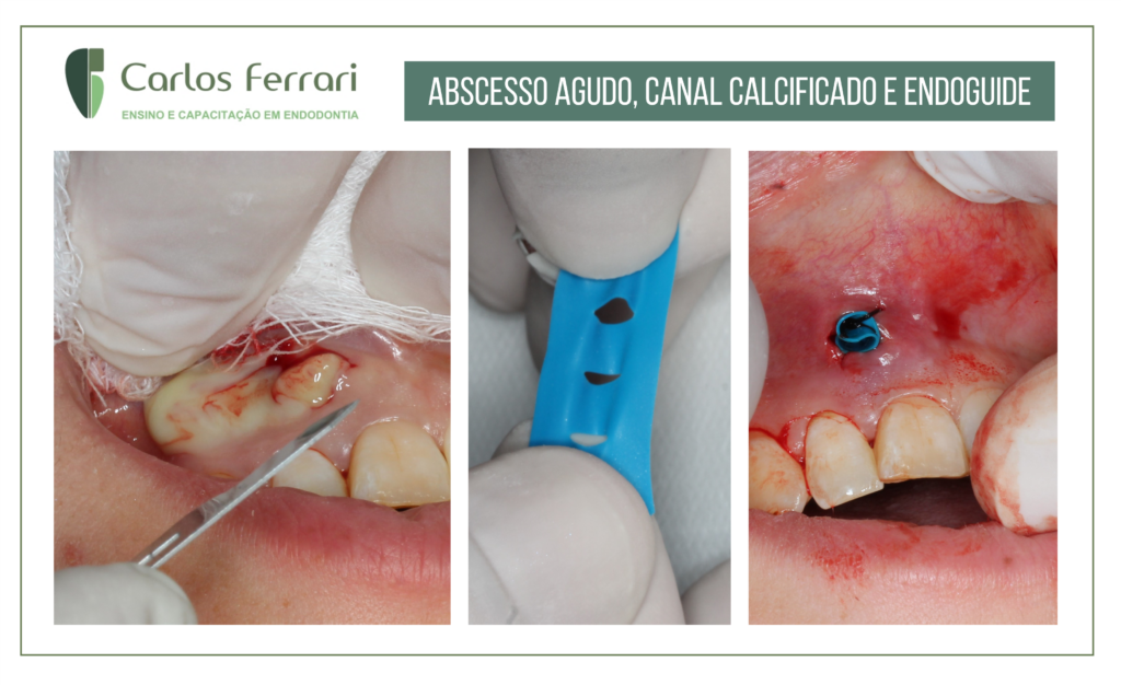
Caso de abscesso periapical agudo. Paciente procurou o consultório queixando-se de dor e inflamação na gengiva na região superior. No exame clínico, observou-se edema com ponto de flutuação na região gengival apical do dente 11, além de palpação e percussão positivos. No exame radiográfico, constatou-se a obliteração total da câmara pulpar. Decidiu-se então , como medida urgencial, pela drenagem e manutenção de dreno até que a paciente realizasse exames de tomografia e escaneamento intraoral para confecção de guia de acesso.


in: Taumaturgo, 2018: https://doity.com.br/anais/conexaofametro2017/trabalho/38087
Abscessos Periapicais: diagnóstico e tratamento
Introdução – A região periapical é uma estrutura dinâmica composta por tecidos que apoiam e envolvem o dente. Esses tecidos incluem o ligamento periodontal, o cemento e o osso alveolar. Os suprimentos vasculares e o nervoso dos tecidos também são vitais ao funcionamento normal dos tecidos periodontais. As alterações periapicais podem ocorrer no organismo humano, em decorrência de reações inflamatórias agudas e crônicas nestes tecidos. Com características etiopatogênicas, clínicas, radiográficas e microscópicas, exclusivas e próprias da região periapical, o abscesso dentoalveolar trata-se de uma coleção purulenta proveniente da morte de células de defesa(neutrófilos, macrófagos) e restos de bacterias, ocasionando sintomatologia dolorosa no paciente. O estado patológico já pode ser detectável através dos exames anamnésico-clínico-radiográfico indicando assim a escolha do tratamento conservador ou cirúrgico.
O diagnóstico dos abscessos possuem diferentes níveis de evolução, devendo ser correto para determinar o tratamento mais adequeado através de uma conduta anamnésico-clinico-radiográfico própria para cada fase do abscesso. Divide-se as fases dos abscessos periapicais em aguda e crônica. No abscesso agudo, a coleção permanece confinado à região periapical, neste caso, o paciente apresenta dor contínua, localizada e pulsátil, sendo detectável uma sintomatologia dolorosa nos exames à palpação apical e percussão. As características radiográficas apresentam quase ou nenhuma alteração, na região do espaço periodontal encontra-se normal ou levemente aumentado e interrupção da lâmina dura. Na continuidade do processo, a evolução do abscesso se difunde para região intraperiosteal (fase óssea, subperiosteal e flegmatosa), o pus tenta perfurar a cortical óssea e se acumula sob o periósteo, o que caracteriza a fase subperiosteal, evidenciando sinais de edema na região acometida de consistência dura, caracterizando abscesso dentoalveolar em evolução. O estágio mais avançado, abscesso dentoalveolar evoluído, apresenta sinais de edemas com pontos de flutuação do material purulento. Quando consegue exteriorizar-se, forma o que se denomina fístula. Com isso, a diminuição ou desaparecimento da dor é praticamente instantâneo, sendo necessário no exame anamnésico identificar presença de sintomas sistêmicos de febre já sendo necessário o manejo de antibioticoterapia. Se este quadro clínico não for resolvido, com o tempo se torna um abscesso crônico, caracterizado por um processo supurativo instalado na região do periápice dental. Como causa constitui basicamente da presença de bactérias na região periapical o tratamento é basicamente a eliminação ou redução da população bacteriana. Uma drenagem cirírgica se faz necessário na maioria dos casos, devido a presença de dor. A escolha do tratamento é determinada pela severidade de sinais e sintomas.
Considerações finais: Portanto, o reconhecimento precoce dos aspectos clínicos e conhecimento da localização da coleção purulenta, durante o processo dinâmico de disseminação, é fundamental para que se possa estabelecer um adequado plano de tratamento.
https://ferrariendodontia.com.br/endoguide-incisivo-abscesso/


