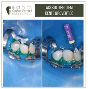
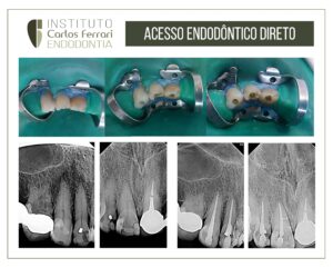
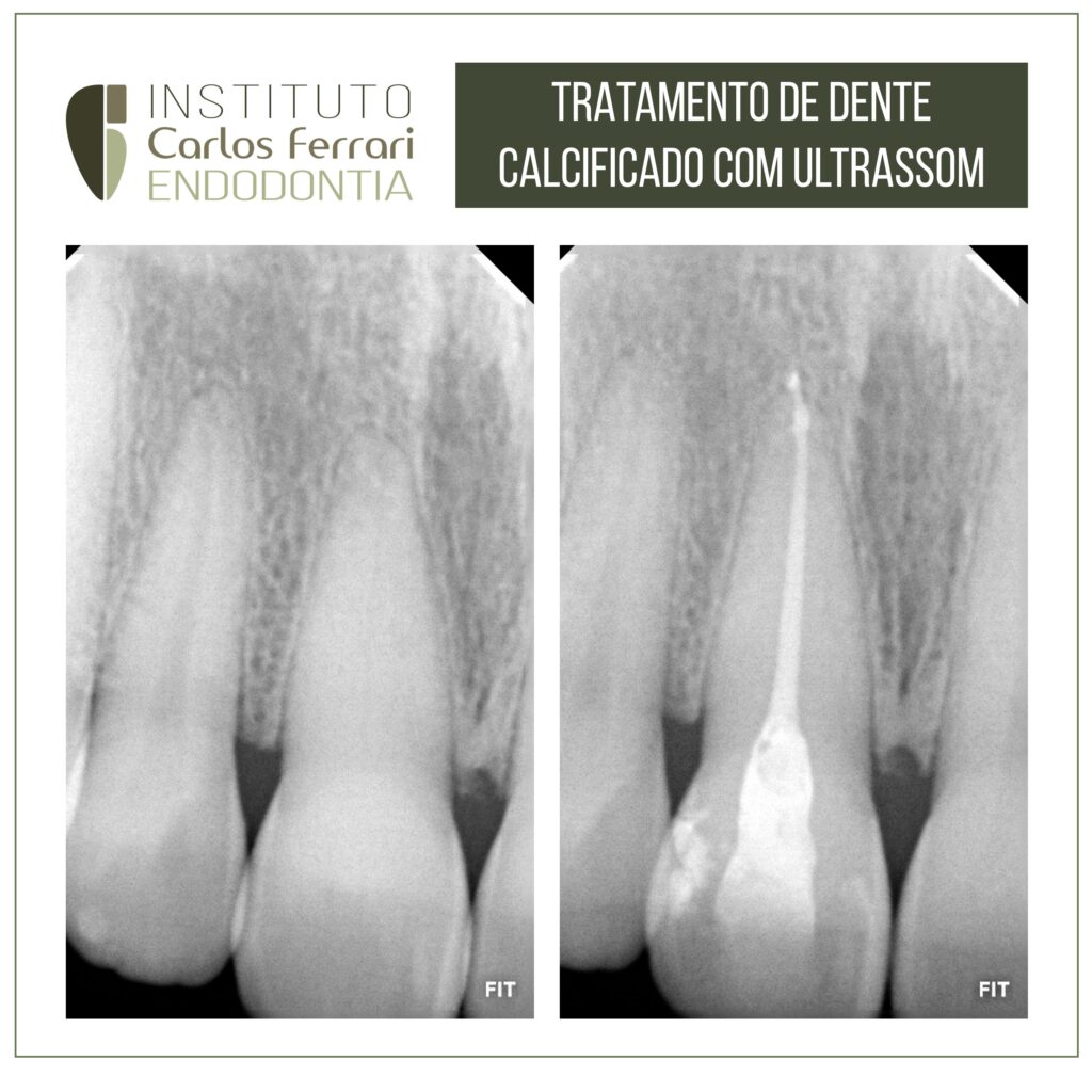
Canal calcificado e ulltrassom. Exploração e tratamento com pontas ultrassônicas.
A localização de canais calcificados apresenta dificuldades que muitas vezes nos levam à utilização de recursos como Endoguide ou mesmo a cirurgia paraendodôntica.
Porém uma exploração realizada com paciência e instrumentos adequados, pode levar ao sucesso.
Nesse casos foi realizada a exploração com instrumentos finos e pontas de ultrassom, até a localização do canal e consequente tratamento.
Caso realizado pelo aluno Igor Fajan, de especialização em endodontia da APCD Bragança Pta.
In: Ribeiro & Aguiar. Revista Ibero- Americana de Humanidades, Ciências e Educação- REAS
INTRODUÇÃO
Na anatomia dentária é indispensável abordar sobre o complexo dentino pulpar, sendo esse uma região suscetível a sofrer danos, capazes de gerar obliteração dos canais radiculares, este fenômeno é causado por diversos fatores, como cáries, envelhecimentos,
traumas, dentre outros que podem atingir a estrutura dentária. Fisiologicamente, os odontoblastos passam a depositar uma quantidade de dentina secundária (em casos de idade, onde é depositada lentamente com o tempo) ou terciária (caso se trate de alguma lesão ou trauma), sendo esta última mais desorganizada e depositada mais rapidamente. As massas de tecido calcificado no nível da câmara pulpar e canais radiculares do dente são denominados de Calcificação Pulpar (CP). (BUCHGREITS J. 2016), (NEVILLE B.ALLEN C. 2004), (SATHEESHKUMARP. 2013).
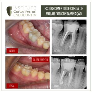
A CP pode estar presente no nível da câmara pulpar e/ou canal radicular.³ podendo até obliterar o canal radicular completamente e esta não pressupõe obrigatoriamente alterações na cor da coroa do dente, no periápice do dente e na sensibilidade do dente. Podem ocorrer em dentes saudáveis, doentes, irrompidos ou impactados em todas as faixas
etárias 6. (SATHEESHKUMAR P. 2013), (MCCABE P. DUMMER P. 2012), (TASSOKER M., MAGAT G. SENNER S. 2018).
As calcificações podem ser localizadas mais regularmente na porção coronária do que na porção radicular do órgão pulpar. Asocorrências relatadas divergem de 8% a 90%,mas diferentes pesquisadores registraram a prevalência em quase 20% de dentes
registrados individualmente por meio de radiografias. Nas calcificações pulpares asprevalências, parecem aumentar com a idade dos indivíduos devido a inúmera quantidadede impactos físicos e químicos, aos quais os dentes são constantemente expostos ao longoda vida dos indivíduos, até chegar à senilidade.(RAVANSHAD S. 2015).
Canal calcificado.