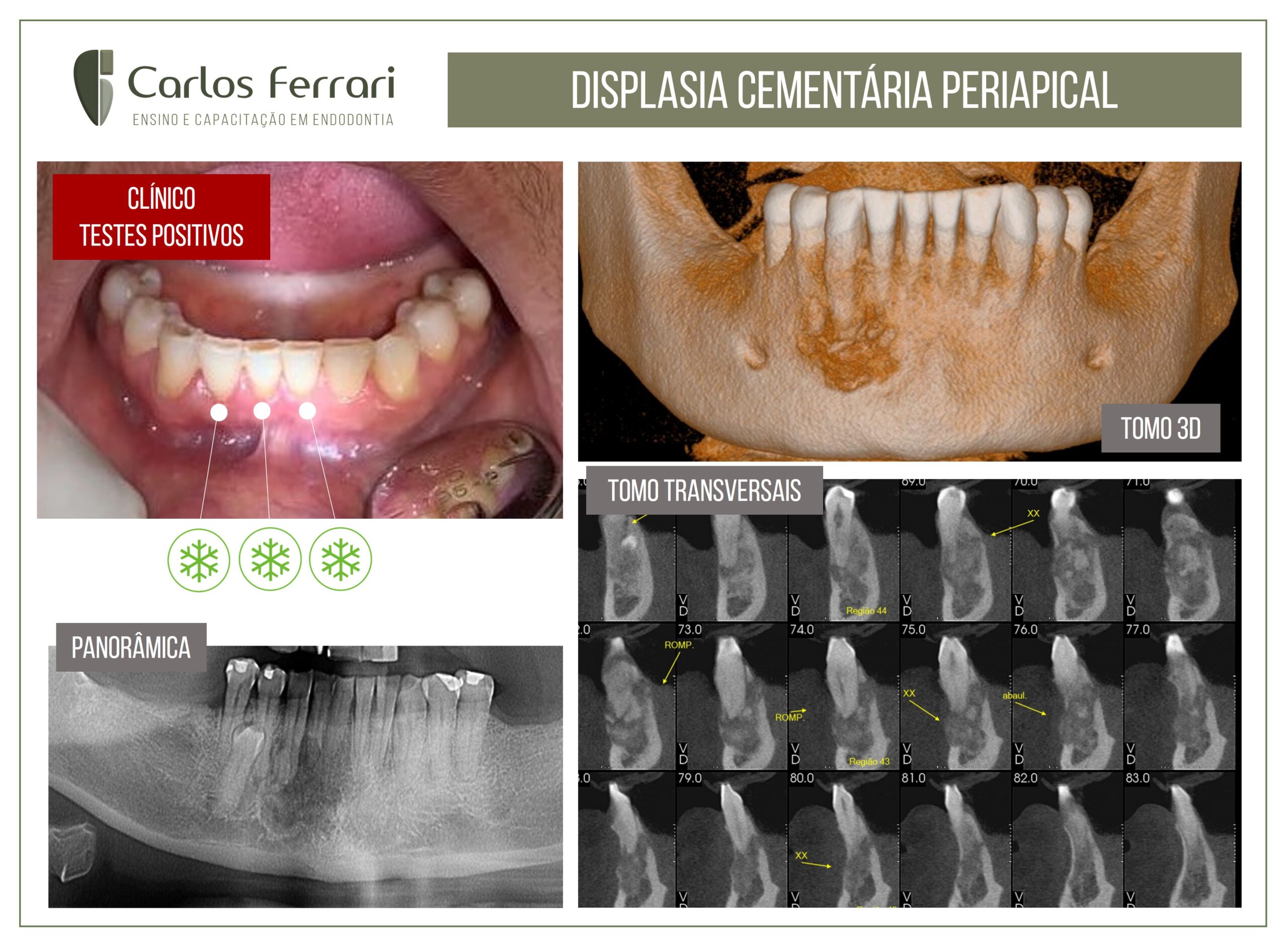
Case of periapical cemental dysplasia. A 46-year-old black female patient was sent for endodontic evaluation due to a radiolucent periapical image in the region of tooth 43. On clinical examination, normal gingival tissues in the region. In the tests, absence of pain on palpation and percussion and normal pulp vitality in this and adjacent teeth. Radiographic examination showed a mixed image in the periapical region from tooth 43 to tooth 44. The diagnostic hypothesis was then of periapical cemental dysplasia, a benign lesion that does not require any intervention. The patient was then advised to return for follow-up and follow-up.
Case conducted by student Cristiane Nestor, from the endodontics specialization course at HPG Brasília.
Periapical cemental dysplasia (PCD) is a benign odontogenic tumor of mesenchymal origin, derived from the periodontal ligament (BADEN, 1987). Its etiology still remains unknown, although some authors believe that it represents an unusual reaction of the periapical bone to a local irritant factor (NEVILLE, 2004); while others suggest that trauma, hormonal disorders, systemic and genetic factors may be involved in its development (SMEELE, 1991).
There are no clinical manifestations and are therefore asymptomatic, except when located near the mental foramen, due to nerve compression that can lead to pain and paresthesia. For these reasons, this lesion is usually discovered in routine radiographic examinations. There is a marked predilection for women (14:1) and approximately 70% of cases affect blacks. The diagnosis is never made before the age of 20. On imaging examinations they are most frequently seen in areas corresponding to the roots of the lower incisors. Multiple foci are more usual, but when single, they do not exceed 1 cm in diameter. It can also be seen that the lesion is contiguous to the periodontal ligament and the lamina dura remains intact (SMITH, 1998).
This pathology has a natural evolution, going through three phases of development: osteolytic; cementoblastic and maturation. The 1st phase is characterized by the replacement of bone tissue by fibrous tissue, which determines a periapical radiolucency. In the 2nd phase, there is deposition of cementum spicules due to increased cementoblastic activity, causing areas of opaque focal condensation with areas of rarefaction. In the final, maturing phase, there is complete calcification of the region providing a radiopaque image, often surrounded by a narrow radiolucent halo
Knowing the characteristics of PCD, its great similarity with other pathologies such as those of endodontic origin (cysts and granuloma) can be noted, and in this regard, the correct diagnosis and performance of the pulp vitality test is essential, since in cases of PCD the pulp is sensitive to the test, not justifying unnecessary endodontic intervention.
https://ferrariendodontia.com.br/lesao-periapical-dens-invaginatus/