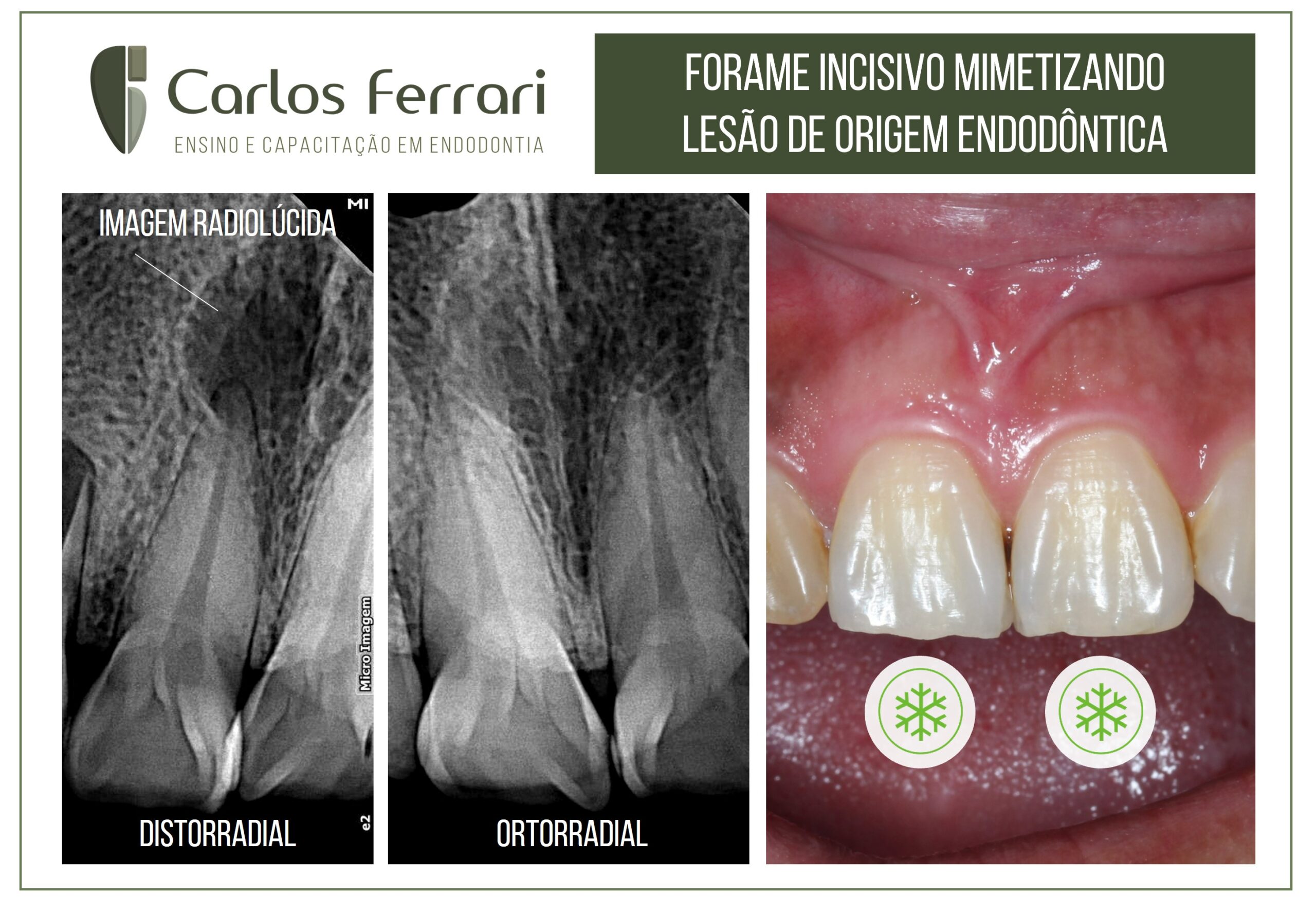
Female, 40 years old, came to the clinic with an indication of treatment of periapical lesion in tooth 11 by observation of circumscribed radiolucent periapical image. On clinical examination, normal aspect of the crown in relation to color and tooth 21, besides positive and normal response to thermal test, percussion and palpation. The periapical radiographic examination revealed that with the change of the horizontal angulation of the x-ray beam, the image "disappeared" from the periapex, leading to the conclusion that it was an overlapping image of the incisive foramen.
Many periapical images confuse the dental surgeon and may be interpreted as a periapical lesion of endodontic origin. Every care should always be taken with regard to careful clinical and radiographic examination, following all the steps to reach a diagnosis. Multiple radiographic views and application of Clark's technique is fundamental in cases like this.
Case conducted by students Karlon Costa and Robson Ribeiro, from class VII of the specialization in endodontics at HPG Brasília.
https://ferrariendodontia.com.br/livro-endodontia-ricardo/
https://www.youtube.com/watch?v=QMLd1CcjsO0&t=1130s
Excerpt from the chapter: Radiology Applied to Endodontics. Ferrari et al, in Machado R. Endodontia. Bases Biológicase técnicas. Ed. Gen, 2002.
Overlapping images in Endodontics
Some anatomical structures present radiolucent radiographic images that, depending on their position, can be confused with periapical bone rarefaction of endodontic origin. Variations in X-ray beam angulation (Clark technique), together with previous information obtained through anamnesis and clinical examination, are fundamental for the differential diagnosis.
In the maxilla, the structures capable of producing such effect are the palatine canal and foramen, which can have their image superimposed on the apexes of the upper central incisors, especially in radiographs with distroradial incidence. Another anatomical structure frequently confused with periapical lesions in premolars and especially in maxillary molars is the maxillary sinus (Figure 6.20). Its anatomical and morphological heterogeneity can produce images similar to periapical lesions that can confuse even experienced clinicians. Also in the maxilla, the periodontal ligament adjacent to the molar roots can simulate the presence of root fractures and additional canals (Figure 6.21).
In the mandible, the anatomical structures that most cause doubts in the interpretation of radiographic exams performed by different techniques are the mandibular canal - which overlaps the root apices of the molars - and the mental foramen - which can mimic a periapical lesion in premolars (Figure 6.22).
Non-endodontic pathologies with periapical radiographic manifestations
Periapical pathologies or changes not caused by pulpoperirradicular involvement may present images often confused as such. A thorough examination of the patient, initially considering the initial complaint, the case history and the results of a thorough clinical examination can in most cases provide data that allow a correct diagnosis. The absence of signs or symptoms and normal responses to vitality tests, percussion and palpation in all teeth adjacent to the lesion can already constitute a substantial indication of its non-endodontic origin.
Radiographic interpretation must be performed with attention and rigor, following specific and objective criteria, among which are: number of lesions, location, radiolucency, border characteristics, internal structures, and effects on adjacent structures and teeth.31 Such questions, when analyzed in order, facilitate the determination of diagnostic hypotheses.
The pathologies and changes of non-endodontic origin most commonly misdiagnosed as such are described below, according to their radiolucency.
Radiopaque
-Idiopathic osteosclerosis: a single image, usually present in the periapical region of the lower posterior teeth, with variable radiopacity, irregular borders and absence of cortical bone (Figure 6.26). It has no known etiology and requires no treatment, being frequently confused with condensing osteitis, caused by a chronic inflammatory process of endodontic origin
-Mandibular torus: external bony formations located on the mandibular lingual surface can produce radiopaque images on periapical examination with variable number and rounded shape, often overlapping the periapical region of the lower teeth (Figure 6.27). The clinical examination can confirm the suspicion by observing such formations through intraoral inspection.
Mixed
Cemento-osseous dysplasias: idiopathic lesions in which there is replacement of bone tissue by fibrous tissue. They present a mixed image, being more radiolucent in the first phase, becoming mixed and then radiopaque after maturation. They are more common in black patients, females and in adulthood, self-limiting and frequently diagnosed by means of routine radiographs. However, because they are located in the periapical region, their presence is commonly associated with unnecessary endodontic treatment. Cemento-osseous dysplasias are subdivided into periapical cemento-osseous dysplasia, more localized in only one region (often in the anteroinferior teeth), and florid cemento-osseous dysplasia, with multiple images spread throughout the arch (Figure 6.28)
-Other pathologies with mixed radiographic images that can mimic lesions of endodontic origin are odontomas and benign cementoblastoma.
Radiolucent
-Cysts: Several cysts of non-endodontic origin are capable of producing images similar to those originating from pulpo-perirradicular involvement, such as: the lateral periodontal cyst (usually located in the region of lower premolars); the dentigerous cyst (more frequent in lower molars) (Figure 6.29), the keratocyst (common in the mandible) (Figure 6.30), the traumatic bone cyst and the nasopalatine cyst (frequent in maxillary anterior teeth)
-Other pathologies capable of mimicking periradicular lesions are: adenomatoid odontogenic tumor (most common among lateral incisors and maxillary canines), giant cell lesion, intraosseous fibromas and lipomas, ameloblastoma, and malignant tumors. An anatomical alteration that produces a radiolucent image in the mandibular angle region, but which can rarely appear in the periapical region, is Stafne's bone defect.