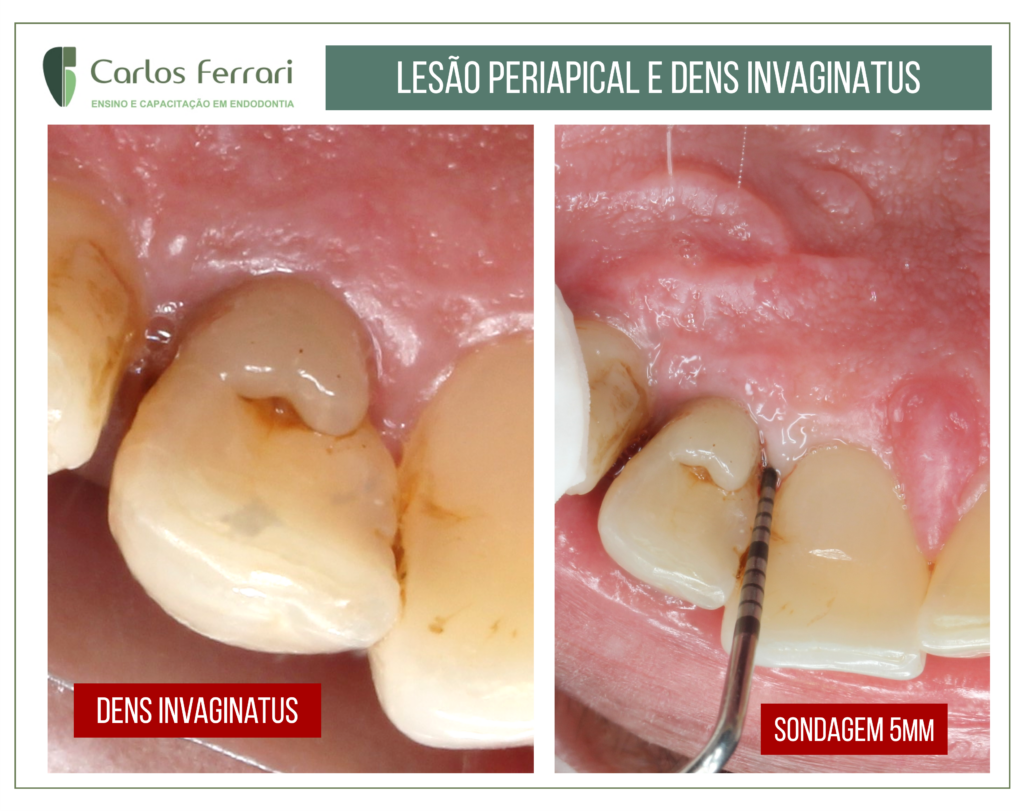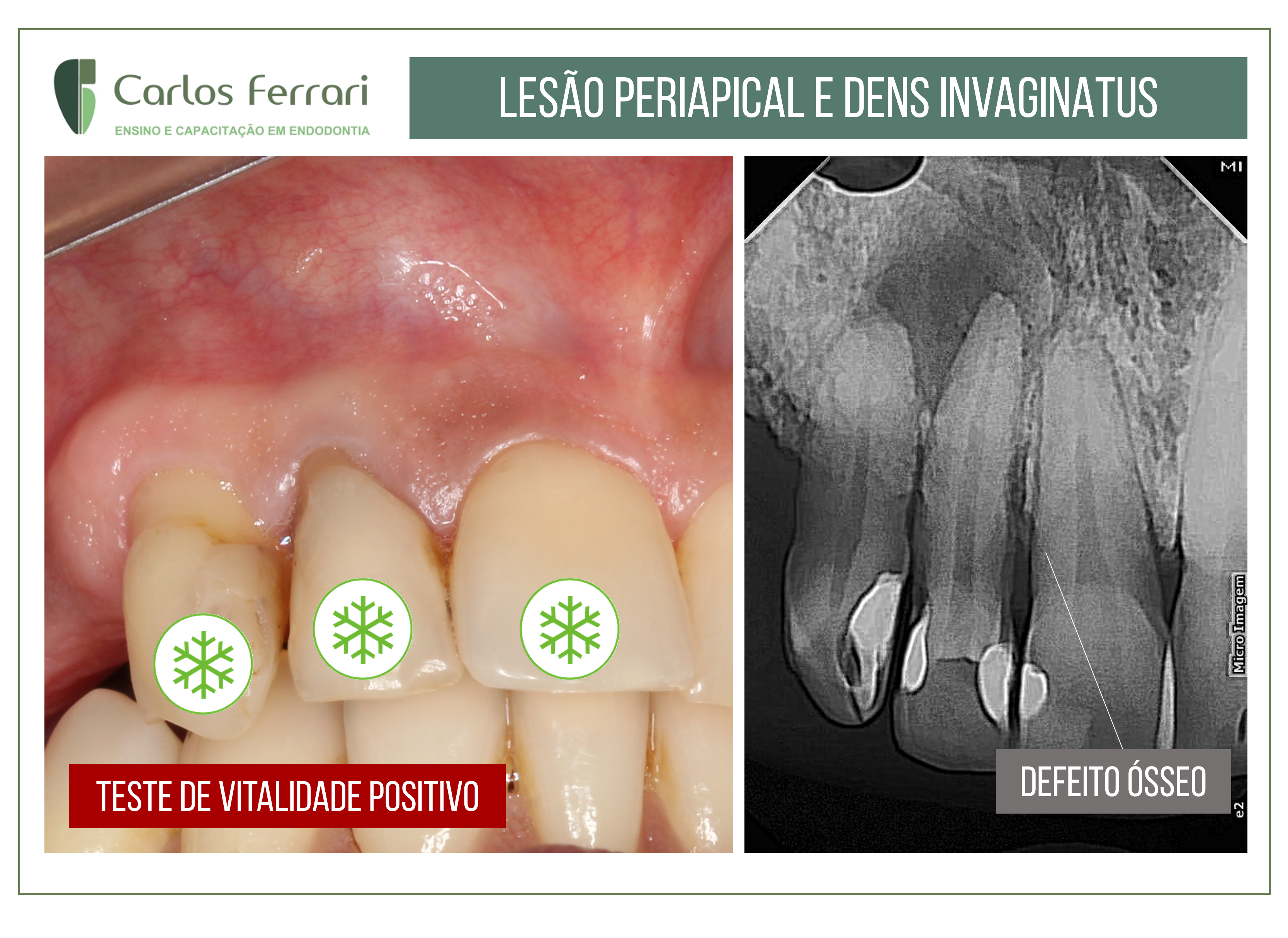
Paciente encaminhado por colega para diagnóstico e conduta devido à uma imagem radiográfica radiolúcida da região anterior direita. Paciente assintomático. No exame clínico, observou-se má formação na região mesial do dente 12 e sondagem periodontal positiva de 5 mm na mesma região. O teste de vitalidade revelou respostas normais para os dentes 11, 12 e 13. No exame radiográfico, imagem radiolúcida periapical na região do dente 22 e imagem de defeito ósseo na região mesial periradicular do dente 12. O caso alerta para a necessidade de um exame mais detalhado para evitar diagnósticos errados em caso de lesões periapicais. A paciente foi indicada para o exame tomográfico e uma melhor observação da área permitirá um estabelecimento mais acurado de uma hipótese de diagnóstico, e só assim um tratamento poderá ser estabelecido.
Caso conduzido pelos alunos Carlos e Vinícius da Especialização em Endodontia da HPG Brasília.