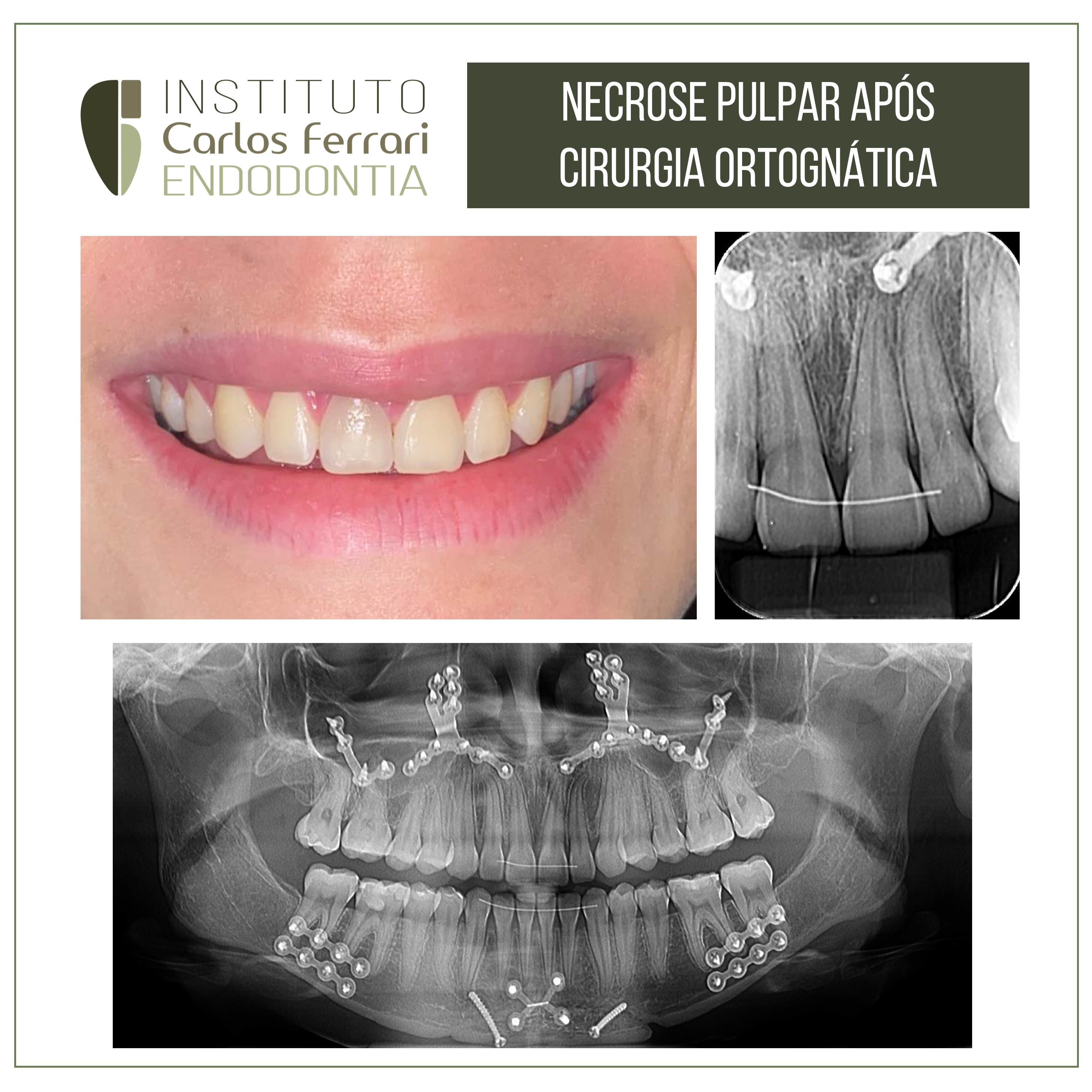
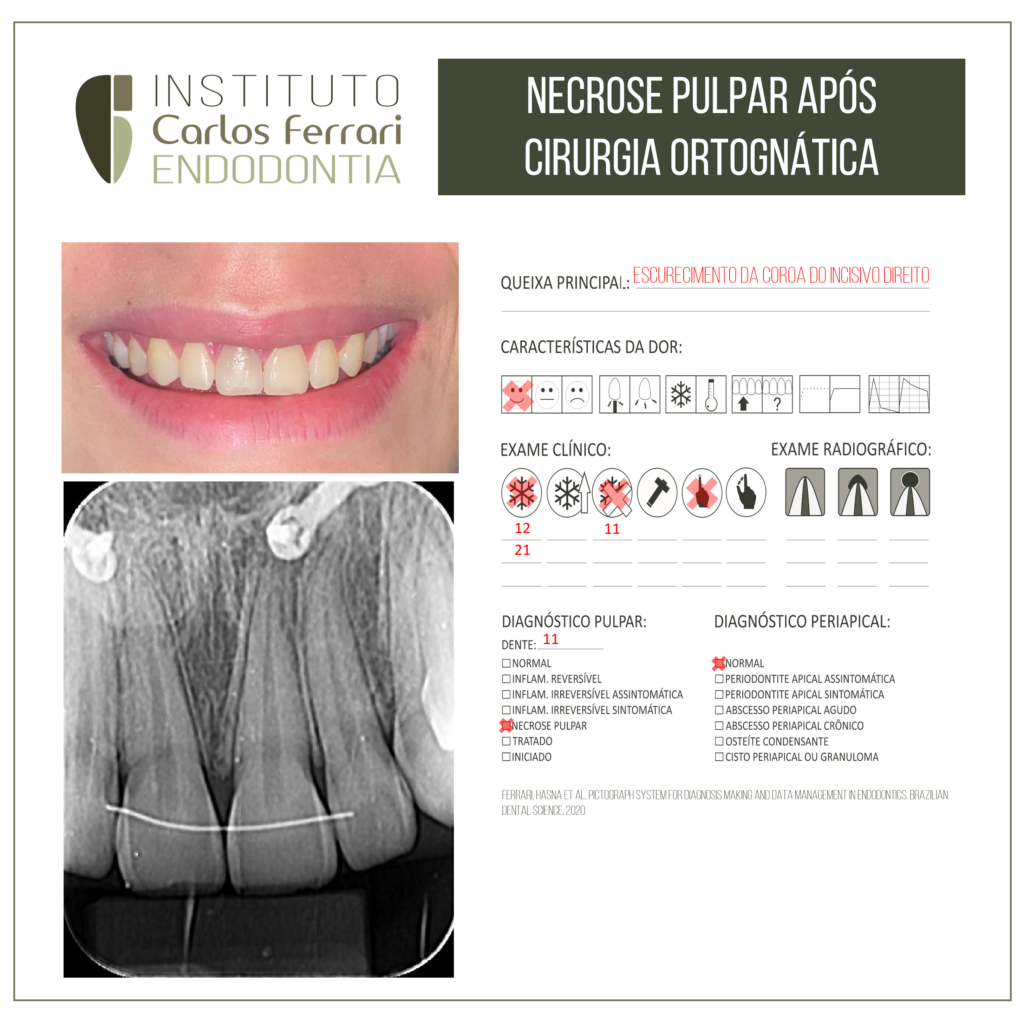
Necrose pulpar após cirurgia ortognática. Paciente procurou atendimento queixando-se de escurecimento da coroa do incisivo central.
In Consolaro et al. Escurecimento dentário e necrose pulpar após cirurgia ortognática: o laringoscópio e o traumatismo dentário. R Dental Press Ortodon Ortop Facial 16, v. 12, n. 5, p. 16-19, set./out. 2007.
A maioria dos trabalhos sobre os efeitos da cirurgia ortognática na polpa dentária compara e ressalta que os efeitos são os mesmos encontrados em dentes submetidos ao traumatismo dentário. Estes trabalhos analisam casuísticas onde foi utilizada a
osteotomia por Le Fort I, com ou sem segmentação, ou ainda da osteotomia mandibular.
Ainda sobre os trabalhos, a grande maioria mediu as alterações do fluxo sanguíneo pulpar utilizando a fluxometria por laser Doppler7 . Raramente foram realizadas análises microscópicas e imaginológicas metodologicamente bem definidas e inquestionáveis. Outra metodologia muito empregada foi o eletrodiagnóstico, para detectar a sensibilidade pulpar.
A precisão diagnóstica e a praticidade da fluxometria pulpar via laser Doppler são questionadas como metodologia mais adequada para análise de viabilidade pulpar, considerando-se que foi idealizada para tecidos moles. A presença de tecido
duro poderia comprometer a sua efetividade e a precisão de resultados. Da mesma forma, são questináveis resultados obtidos apenas pelo eletrodiagnóstico pulpar.
Alguns estudos preocuparam-se em analisar a ocorrência da necrose pulpar, do escurecimento dentário e da reabsorção interna, independentemente se em dentes anteriores ou posteriores, mas há uma concentração de atenção para com os incisivos centrais superiores.
Os dentes traumatizados podem: a) manter sua vitalidade pulpar; b) sofrer necrose pulpar asséptica; c) sofrer metamorfose cálcica da polpa.
A necrose pulpar asséptica pode evoluir gradativamente para o escurecimento dentário e este sinal clínico ser a indicação de que algo está ocorrendo com o dente portador. Este sinal pode ser notado pelo paciente ou pelo profissional, ou ambos, simultaneamente. A metamorfose cálcica da polpa foi muito conhecida clinicamente como obliteração da câmara ou do canal radicular, comumente vista em dentes traumatizados, após algumas semanas ou meses.
A metamorfose cálcica da polpa ocorre quando o traumatismo lesa o feixe vásculo-nervoso, não o suficiente para impedir o fluxo sanguíneo, mas sim restringi-lo. Nestas condições de sofrimento pulpar ou estresse celular intenso por hipóxia, as células da polpa sofrem uma metaplasia, ou seja, mudam seu fenótipo e todos os fibroblastos, pe
ricitos e células de reserva diferenciam-se aleatoriamente em odontoblastos e, de forma desorganizada, depositam uma matriz dentinária displásica no meio extracelular, ou seja uma dentina mal formada, cheias de células incluídas, a ponto de ser denominada de osteodentina.
A metamorfose cálcica da polpa pode ser simultânea em toda a polpa, mas pode ser detectada imaginologicamente ocorrendo das paredes para o centro da polpa, reduzindo gradativamente o volume pulpar a ponto de, em 6 a 12 meses, o canal estar completamente obstruído ou estreitado. Não raramente ocorre a obliteração da câmara pulpar e o estreitamento do canal radicular.
Necrose pulpar após cirurgia ortognática.