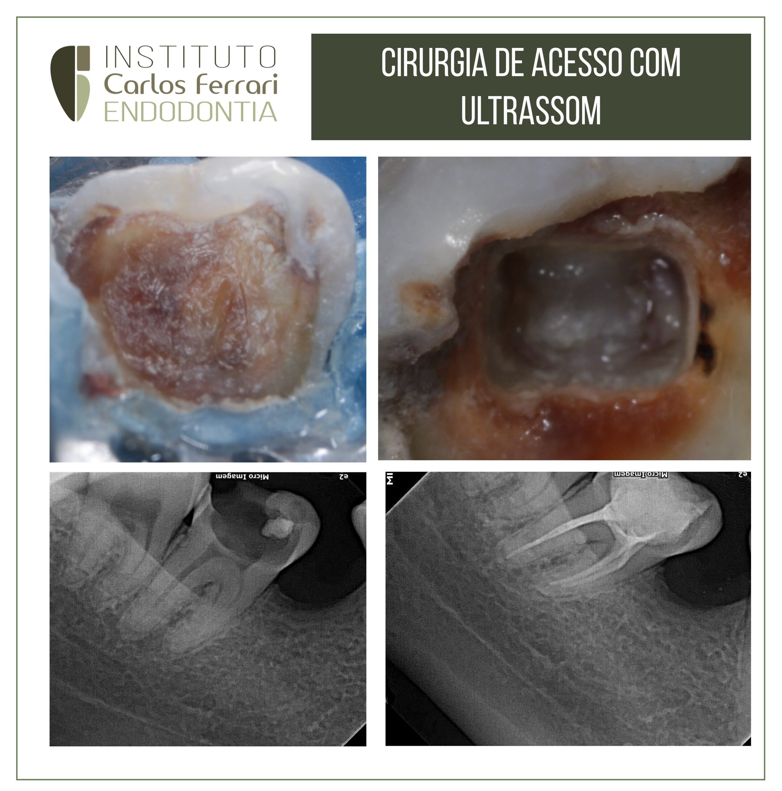
CPonta de ultrassom na endodontia
Utilização de ponta de ultrassom, em potência média para remoção de dentina terciária acumulada na câmara pulpar de um molar inferior.
Caso realizado pelos alunos Andressa Camargo e Igor Fajan, da especialização de endodontia da APCD Bragança Pta.

In: Figueiredo et al. Magnificação e ultrassom como recursos auxiliares no tratamento endodôntico em dentes com calcificação: considerações clínicas e relato de caso. Arch Health Invest (2021) 10(1):174-178
INTRODUÇÃO
Os objetivos do tratamento endodôntico (TE) consistem na limpeza, modelagem e preenchimento através da obturação tridimensional do sistema de canais radiculares (SCRs), estando o sucesso a longo prazo do TE intimamente associado a tais aspectos.
Entretanto, para a obtenção do sucesso etapas operatórias como a adequada realização da cirurgia de acesso e consequentemente a localização e identificação da abertura do canal radicular são requisitos importantes.
Uma dificuldade que pode estar presente durante o TE consiste na obstrução do orifício da entrada dos canais radiculares, o qual pode ocorrer em virtude da presença de estruturas calcificadas sob a forma de depósitos de dentina secundária ou de calcificações pulpares (CPs) propriamente ditas.
A presença de estruturas calcificadas na polpa dental são alterações bastante comuns, podendo afetar dentes decíduos ou permanentes. Enquanto estruturas calcificadas decorrentes da deposição de dentina secundária podem ocorrer em virtude da
resposta à presença de material restaurador inserido próximo à polpa, outras CPs podem ocorrer livremente na polpa ou estarem aderidas às paredes dentinárias, apresentando-se sob a forma de calcificações difusas ou de nódulos pulpares (NPs)3,5, podendo os NPs apresentarem formato oval ou irregular de tamanho variável.
Tanto a deposição de dentina secundária quanto as CPs podem obliterar a câmara pulpar. Consequentemente, tanto esta, quanto as CPs3 podem obstruir os orifícios de entrada dos canais e dificultar a localização e o acesso ao SCRs.
A tomografia computadorizada de feixe cônico (TCFC) tem se tornado uma ferramenta imprescindível na prática clínica endodôntica durante o diagnóstico e o planejamento do TE , destacando-se como importante método auxiliar na avaliação da anatomia do SCRs bem como no diagnóstico de CPs e dalocalização de canais calcificados.
Além disso, o uso de recursos auxiliares como o microscópio operatório (MO) promove uma magnificação com melhor iluminação e visualização do campo operatório, facilitando a localização dos canais radiculares calcificados e permitindo a execução do TE. Somado a isso, o uso de dispositivos ultrassônicos permitem também auxiliar na localização dos canais radiculares calcificados e/ou de difícil acesso, sendo essas duas estratégias clínicas viáveis para acessar áreas de difícil acesso devido à presença de CPs.
Canal obliterado e ultrassom.
https://ferrariendodontia.com.br/canal-calcificado/
https://www.youtube.com/results?search_query=carlos+ferrari+endodontia+ultrassom