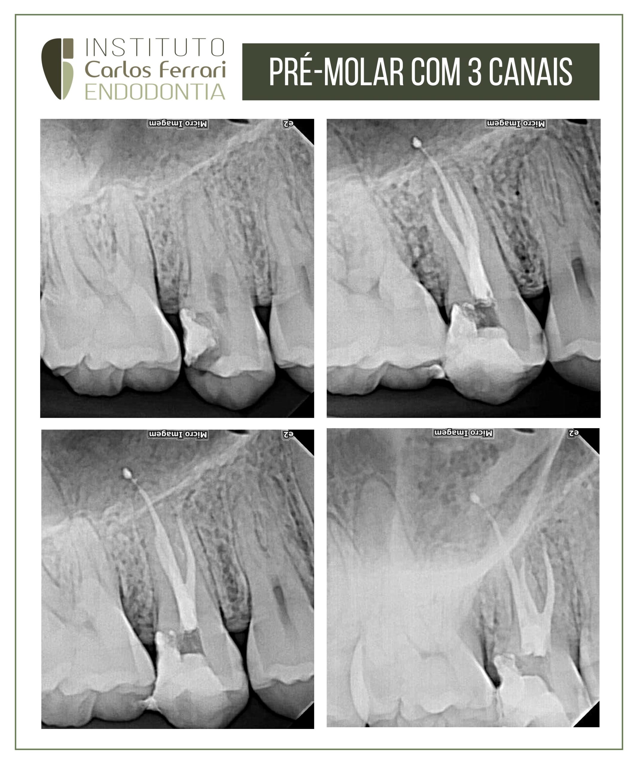
Pré-molar superior com 3 canais. Tratamento endodôntico realizado emsessão única em dente com anatomia com complicações.
In: Machado, Ricardo. Endodontia: Princípios Biológicos e Técnicos. Disponível em: Grupo GEN, Grupo GEN, 2022.
Pré-molares superiores
São os dentes que mais apresentam variações anatômicas considerando o número de raízes e canais (Figura 9.18) e a localização de bifurcações (terço cervical, médio ou apical), as quais são mais comuns nos primeiros (Figura 9.19). Quando da presença de duas raízes/canais, uma será vestibular e a outra palatina. No caso de três raízes/canais, estes serão: mesiovestibular, distovestibular e palatino(a).
Quanto mais apical a bi/trifurcação, mais difícil a localização dos canais. Se o dente apresentar uma raiz, o canal tende a ser amplo no sentido vestibulopalatino e estreito na direção oposta (mesiodistal). Nesses casos, especial atenção deve ser dada à instrumentação, procurando tocar ao máximo todas as paredes. Pré-molares superiores também podem conter istmos54. Estratégias adicionais devem ser adotadas para a limpeza dessas áreas.
Quando há duas raízes, normalmente existem pequenas curvaturas apicais. A porção apical da raiz vestibular é voltada para a palatina e a porção apical da raiz palatina, voltada para a vestibular. Em muitas situações, essas curvaturas não são visualizadas radiograficamente. Considerando-se esse fato e o pequeno volume da raiz vestibular em nível apical, amplitudes de instrumentação exageradas podem provocar perfurações em forma de canaleta.
Pré-molares superiores com raiz única e dois canais poderão apresentar um ou dois forames, fato este associado à forma dos ápices: se o ápice é afilado, os canais tendem a terminar no mesmo forame; se amplo, os canais provavelmente terão forames apicais distintos.
Em 2010, Jayasimha Raj e Mylswamy52 investigaram a morfologia dos canais radiculares de 200 segundos pré-molares superiores extraídos de uma população da Índia, os quais foram corados e diafanizados para a realização de análises estereomicroscópicas. Segundo os resultados do estudo, 64,1% dos dentes apresentavam canais únicos e 35,4% dois canais. O comprimento médio foi de 21,5 mm. Istmos e deltas apicais foram encontrados em 19 e 14% dos dentes, respectivamente.
Em 2012, Zeng et al.57 avaliaram a presença e as características de curvaturas radiculares em 200 primeiros pré-molares superiores de adolescentes de ambos os gêneros (100 homens e 100 mulheres), da província de Guangdong (China). Após a abertura, instrumentos tipo K foram inseridos nos canais radiculares até a saída foraminal. Posteriormente, realizaram-se radiografias periapicais em ambos os sentidos (mesiodistal e vestibulolingual) e os ângulos das curvaturas foram determinados pela aplicação do método de Schneider58. Os resultados do estudo demonstraram que i) 59,21% das raízes eram curvas no sentido vestibulolingual, e estavam presentes em 49,74 e 68,98% dos pacientes do gênero masculino e feminino, respectivamente; ii) 41,05% eram curvas no sentido mesiodistal, estando presentes em 36,27 e 45,99% dos pacientes do gênero masculino e feminino, nessa ordem; iii) 6,84% apresentavam curvaturas em forma de S, das quais 4,15 e 9,63% estavam presentes em dentes de pacientes do gênero masculino e feminino, respectivamente, e; iv) as curvaturas dos canais palatinos de pacientes do gênero feminino foram mais pronunciadas em ambos os sentidos que estas nos mesmos canais de pacientes do gênero masculino. Segundo as conclusões do estudo, canais radiculares curvos em primeiros pré-molares superiores podem ocorrer em ambos os sentidos (mesiodistal e vestibulolingual), ou ainda apresentar dupla curvatura (forma de S). A incidência de canais curvos foi maior em pacientes do gênero feminino.
Al-Ghananeem et al., em 2014 avaliaram o número de raízes e canais radiculares de segundos pré-molares superiores de uma população da Jordânia. Duzentos e dezessete pacientes (100 do gênero feminino [46%] e 117 do masculino [54%]) tiveram seus segundos pré-molares superiores tratados endodonticamente entre janeiro de 2012 e janeiro de 2014. A idade média dos pacientes era de 32,7 anos, variando de 18 a 60 anos. Os dentes incluídos no estudo foram examinados clínica e radiograficamente para análise do número de raízes e canais. Segundo os resultados da pesquisa, 120 e 96 dentes apresentavam uma e duas raízes, respectivamente. Um dente apresentou três raízes. Considerando-se a configuração dos canais radiculares, 30, 54 e 132 dentes apresentavam canais únicos, dois canais terminando em forame único e dois canais com forames apicais distintos, nessa ordem. Um dente apresentou três canais e três forames separados.
Pré-molar superior com 3 canais.