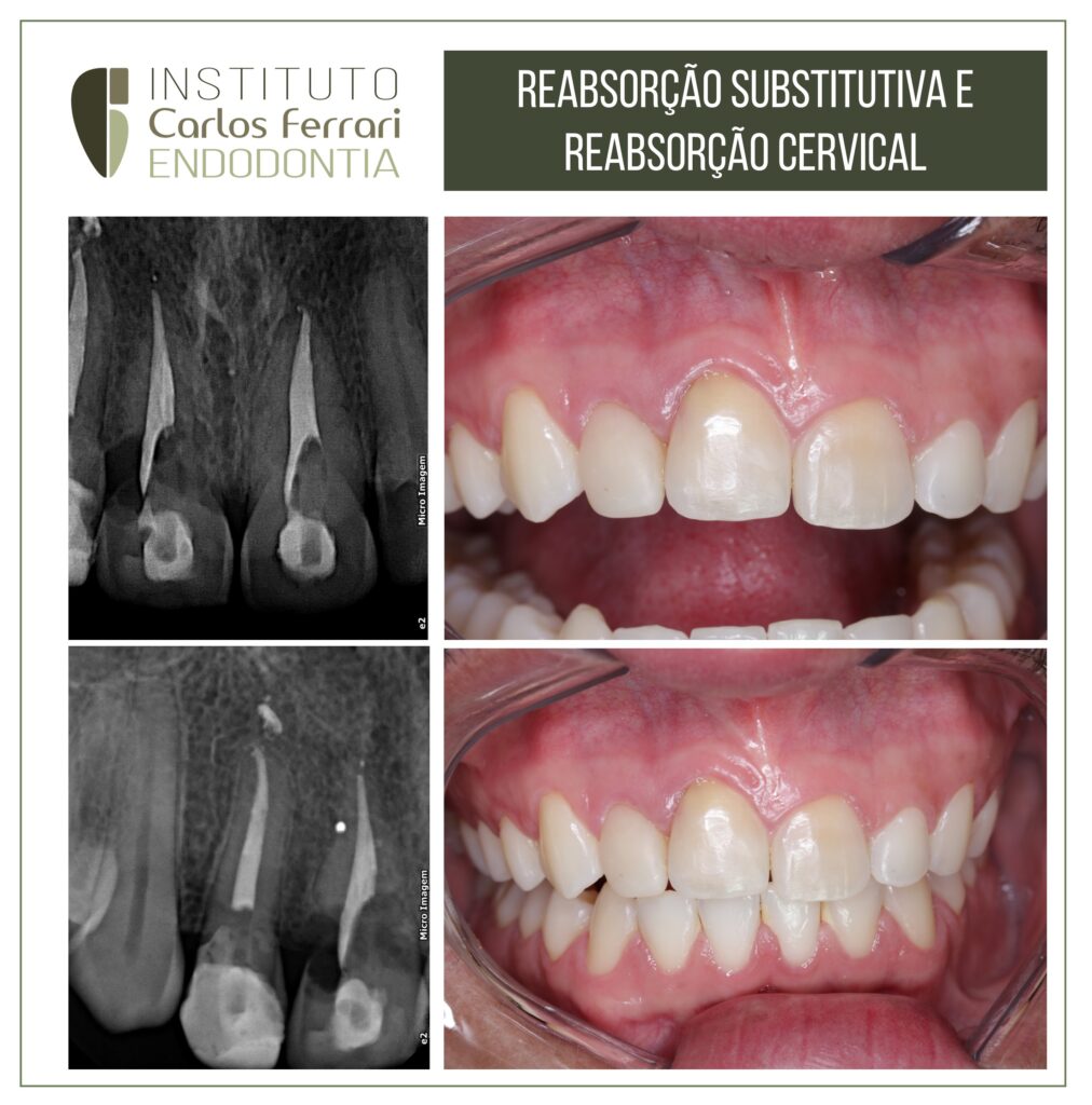
Replacement resorption and cervical resorption.
A 25-year-old patient came to the clinic complaining of pain in the gingival region of tooth 11 and a history of avulsion trauma to the tooth 10 years ago.
Clinical examination revealed a tooth in infraocclusion, a negative mobility test and abnormal percussion tone. Radiographic examination revealed an image compatible with ankylosis (replacement resorption) and cervical resorption. The patient was referred for extraction and implant placement after the prognosis had been explained.
Ankylosis in: Dental Ankylosis. Diagnosis and Literature Review. Journal of the Vale do Rio Verde University v. 17, n. 1: 2019.
Dental ankylosis is considered an abnormality arising from tooth eruption, which occurs in the synthesis of anatomical processes, such as the cementoblastic tissue that covers the roots of dental elements, to the alveolar bone, where the tooth is located, causing an invalidation of the periodontal ligament in some areas around the root (SANTOS et al., 2009).
According to Alves et al. (2011) the impacted tooth may appear impacted, i.e., below the occlusal line or submerged. This change is mistakenly known as "submersion", and it is not the dental element that submerges, but the bone that continues to grow, followed by the soft tissue, which gradually surrounds the ankylosed tooth element.
According to Madeiro et al. (2005), dental ankylosis is considered the most common cause of infraocclusion and its frequency in deciduous teeth ranges from 1.3% to 38.5% in different individuals. The deciduous second molar
is reported as the most affected one. In permanent dentition, its prevalence is lower compared to deciduous teeth. The mandibular third molars are the most ankylosed teeth, followed by the maxillary third molars and canines. In decreasing order of frequency, impaction can also be seen in mandibular premolars, mandibular canines, maxillary premolars and maxillary central incisors.
The pathogenesis of dental ankylosis is unknown and may be secondary to several factors such as disorders arising from changes in local metabolism, trauma, chemical or thermal irritation, failure in local bone development and abnormal tongue pressure (NEVILLE et al., 2004). Dental ankylosis can be classified into three conditions according to the level of infraocclusion, which are: mild, moderate and severe. In the mild condition, the occlusal surface is limited around 1 mm below the occlusal level; in the moderate condition, the occlusal surface is at the level of the contact limit of the contiguous teeth; in the severe condition, the location is at the level or below the interproximal gingival tissue of one or both contiguous dental surfaces (ALVES et al., 2011).
The established therapy for dental ankylosis implies the exodontia of the compromised dental element and its future
replacement by a conventional prosthesis or implant supported, or even the orthodontic closure of the remaining space (VALCANAIA et al., 2003).
Replacement resorption and cervical resorption.