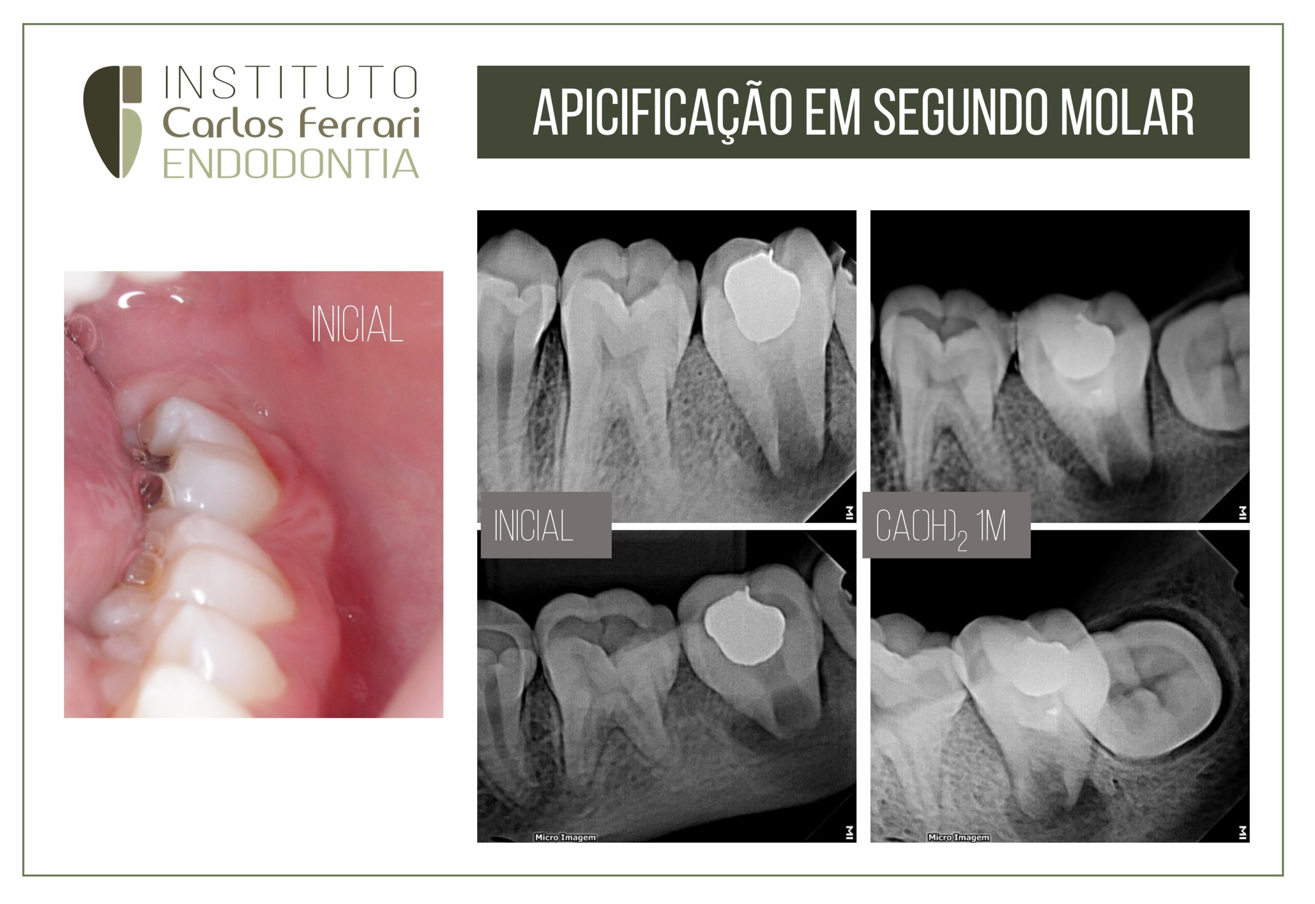
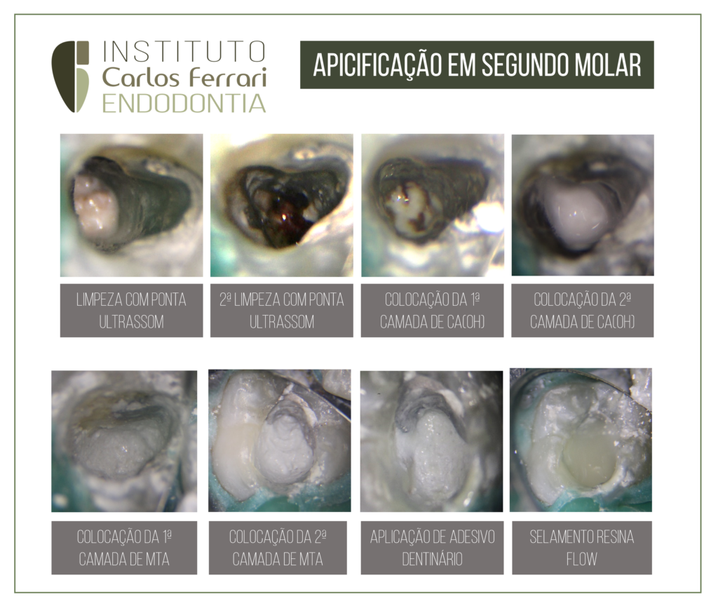
Incomplete rhizogenesis. Apicification in 2nd lower molar with acute periapical abscess.
A 13-year-old male patient was referred by a colleague for endodontic treatment on his lower second molar. He presented pain and swelling in the apical region of the tooth, besides pain on the negative vitality test and positive on percussion and palpation. Radiographic examination showed a diffuse periapical radiolucent image and incomplete root formation, Nolla stage 8.
After decontamination and medication with calcium hydroxide for 30 days, the patient returned with no signs or symptoms to complete the treatment. An attempt was made to induce a clot (revascularization), but after a few minutes, and also due to the patient's behavior, it was considered appropriate to fill with MTA and calcium hydroxide plug.
The patient returned for control after 4 months, in which the continuity of the repair is observed clinically and radiographically. The caregiver was also instructed to complete the restorative treatment.
In: Fernandes et. al. Ciência Atual | Rio de Janeiro | Volume 6, Nº 2 - 2015 | inseer.ibict.br/cafsj | Pg. 02-07:
INTRODUCTION
Traumatic oral injuries followed by orofacial trauma in children and adolescents with young permanent teeth are frequently seen. Most of these incidents occur before complete root
formation and may result in pulpal inflammation or necrosis (RAFTER, 2005). Dental trauma is the
most frequent cause of necrosis in immature permanent anterior teeth. Most cases involved the upper central
incisors, accounting for 70% of traumatized teeth and one third of these teeth had
partially developed root at the time of the accident (SHEEHY et al, 2007).
Apicification is the method of inducing apical closure through the formation of a mineralized tissue
in the apical region of a tooth with necrotic pulp, incomplete root formation and open apex. The procedure requires the chemical-mechanical preparation of the canal, followed by the placement of an intracanal medication to
stimulate healing of the periradicular tissues and the formation of a mineralized apical barrier. The material
most commonly used in apicification is calcium hydroxide. It should not be confused with apicigenesis,
which is also called apicogenesis (treatment of a vital tooth), whose main objective is to stimulate the
physiological development of the root and the formation of the apex (PACE et al.,2007).
Treatment involving teeth with incomplete rhizogenesis requires an accurate diagnosis of the condition of the
dental pulp, and it is essential to define the pathological state of the pulp in order to define the treatment approach for
the tooth.
A thorough clinical and radiographic study will provide important information such as the presence of decayed tissue, fractures, periradicular lesions and stage of root development. For treatment of pulped teeth with incomplete rhizogenesis, after emptying the conduit and cleaning the walls, the canal should be filled with calcium hydroxide-based paste. It is very important that an X-ray be taken to verify that the calcium hydroxide paste has filled the entire root canal. The change of material should be done after
seven days and from then on it can be optional. The larger the foramen opening, the more changes will be necessary.
Furthermore, it must be observed if root closure or development is occurring, which dispenses
the change of calcium hydroxide paste. Among the various substances used for apex closure, the
calcium hydroxide, pure or associated with other substances, has been the material of choice and of greater scientific support
. Clinical and histological studies prove the efficacy of this material over the others. The properties
of calcium hydroxide provide conditions for the tooth to react: it is hemostatic, non-aggressive, and leads the body
to a satisfactory tissue response, stimulating the formation of a mineralized barrier at the cut site,
with biological isolation of the region. (SIQUEIRA et al 2007). Once detected the presence of hard
tissue barrier, through radiographic visualization and clinical inspection all material should be removed from the canal and this
obturated taking care not to exert excessive pressure on the periradicular tissues . The definitive restoration
should be done and the tooth submitted to radiographic follow-up every 6 months.(SOARES et al
2008; LOPES et al, 2010).
