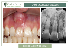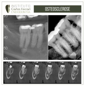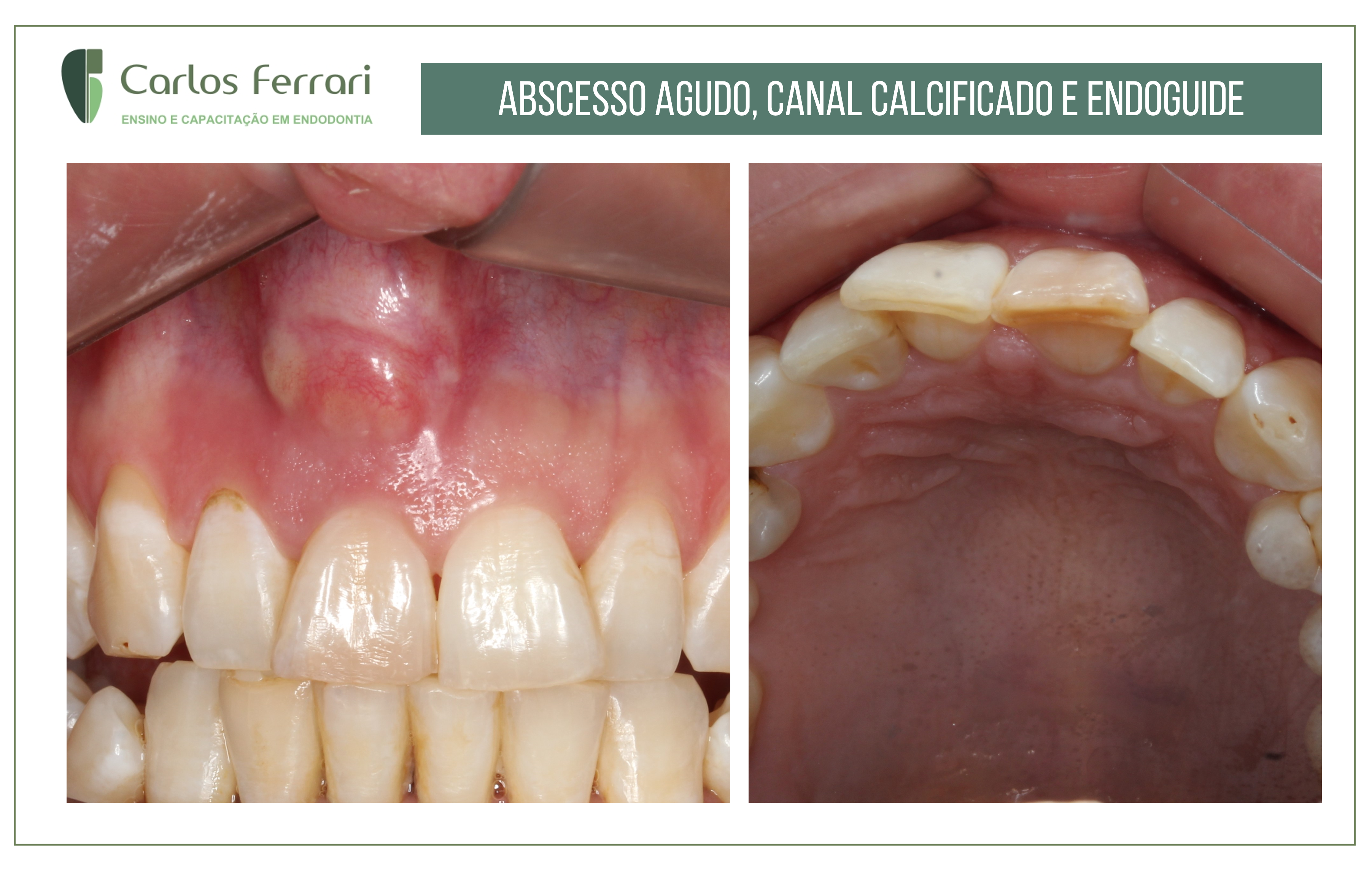
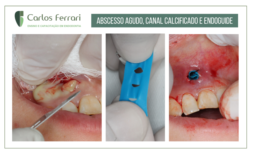
Case of acute periapical abscess. Patient came to the office complaining of pain and inflammation of the gums in the upper region. On clinical examination, swelling with a floating point was observed in the apical gingival region of tooth 11, in addition to positive palpation and percussion. Radiographic examination revealed total obliteration of the pulp chamber. It was decided then, as an urgent measure, to drain and maintain the drain until the patient underwent tomography exams and intraoral scanning to make an access guide.

in: Taumaturgo, 2018: https://doity.com.br/anais/conexaofametro2017/trabalho/38087
Periapical Abscesses: Diagnosis and Treatment
Introduction - The periapical region is a dynamic structure composed of tissues that support and surround the tooth. These tissues include the periodontal ligament, cementum and alveolar bone. The vascular and nervous tissue supplies are also vital to the normal functioning of the periodontal tissues. Periapical changes can occur in the human body as a result of acute and chronic inflammatory reactions in these tissues. With etiopathogenic, clinical, radiographic and microscopic characteristics that are exclusive and specific to the periapical region, the dentoalveolar abscess is a purulent collection originating from the death of defense cells (neutrophils, macrophages) and bacterial remains, causing painful symptoms to the patient. The pathological state can already be detectable through anamnesic-clinical-radiographic exams, thus indicating the choice of conservative or surgical treatment.
The diagnosis of abscesses has different levels of evolution, and should be correct to determine the most appropriate treatment through an anamnestic-clinical-radiographic approach for each phase of the abscess. The phases of periapical abscesses are divided into acute and chronic. In the acute abscess, the collection remains confined to the periapical region; in this case, the patient presents continuous, localized, pulsatile pain, and a painful symptomatology is detectable in examinations by apical palpation and percussion. The radiographic features present almost or no change, in the region of the periodontal space is normal or slightly increased and interruption of the lamina dura. As the process continues, the abscess spreads to the intraperiosteal region (bone, subperiosteal and phlegmatous phases); the pus tries to perforate the cortical bone and accumulates under the periosteum, which characterizes the subperiosteal phase. The most advanced stage, an evolved dentoalveolar abscess, shows signs of edema with fluctuation points of purulent material. When it succeeds in externalizing itself, it forms what is called a fistula. With this, the decrease or disappearance of pain is practically instantaneous, and it is necessary in the anamnestic exam to identify the presence of systemic symptoms of fever, already requiring the management of antibiotic therapy. If this clinical picture is not resolved, with time it becomes a chronic abscess, characterized by a suppurative process installed in the region of the dental periapice. As the cause is basically the presence of bacteria in the periapical region, the treatment is basically to eliminate or reduce the bacterial population. A surgical drainage is necessary in most cases, due to the presence of pain. The choice of treatment is determined by the severity of signs and symptoms.
Final considerations: Therefore, early recognition of the clinical aspects and knowledge of the location of the purulent collection during the dynamic process of dissemination is fundamental in order to establish an adequate treatment plan.
https://ferrariendodontia.com.br/endoguide-incisivo-abscesso/