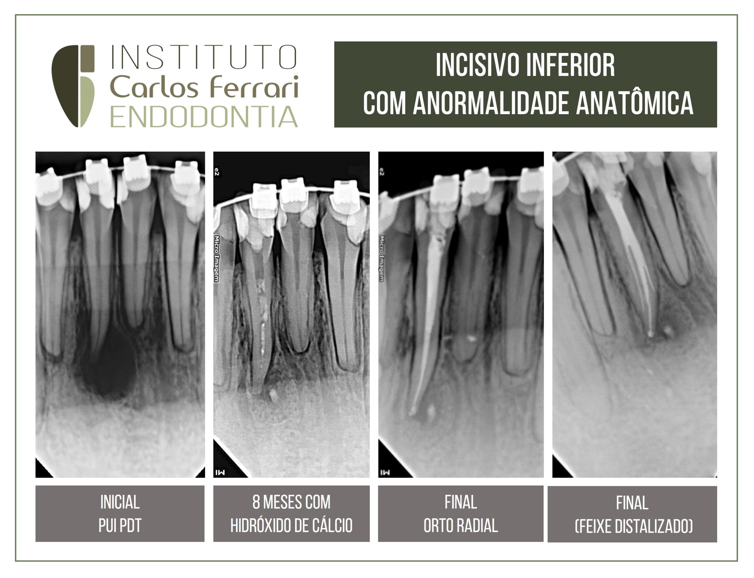
Anatomia dental. Anormalidade anatômica do dente incisivo lateral inferior.
Paciente procurou atendimento para o tratamento endodôntico do dente 41 indicado por outro colega. Não apresentava sinais e sintomas e no exame clínico, dor à palpação e teste térmico negativo no dente 41. No exame radiográfico, imagem radiolúcida circunscrita no referido dente. Verificou-se na exploração dos canais, que o dente tinha mais de um canal. Após o tratamento e medicação com hidróxido de cálcio, o paciente só retornou após 8 meses, e o exame radiográfico revelou imagem de reparação.
Foi realizada então a irrigação e obturação. Observa-se uma anormalidade anatômica, com presença de canais colaterais.
anatomia dentária
In: Machado, Ricardo. Endodontia: Princípios Biológicos e Técnicos. Disponível em: Grupo GEN, Grupo GEN, 2022
Incisivos inferiores
Normalmente, apresentam raízes e canais achatados no sentido mesiodistal, retos ou levemente curvos para distal no terço apical.
Em virtude do achatamento no sentido mesiodistal, canais de incisivos inferiores tendem a ser mais amplos na direção contrária (vestibulolingual). Por esse motivo, a instrumentação não consegue contemplar todas as suas paredes, o que resulta em canais deficientemente instrumentados, limpos e obturados. Ainda, esse achatamento pode predispor à ocorrência de dois canais, um vestibular e um lingual, nem sempre detectados a partir de radiografias convencionais. Em razão de sua raiz ser inclinada para lingual e a câmara pulpar apresentar pequeno volume, incisivos inferiores também são bastante suscetíveis a perfurações durante a abertura coronária.
anatomia dentária