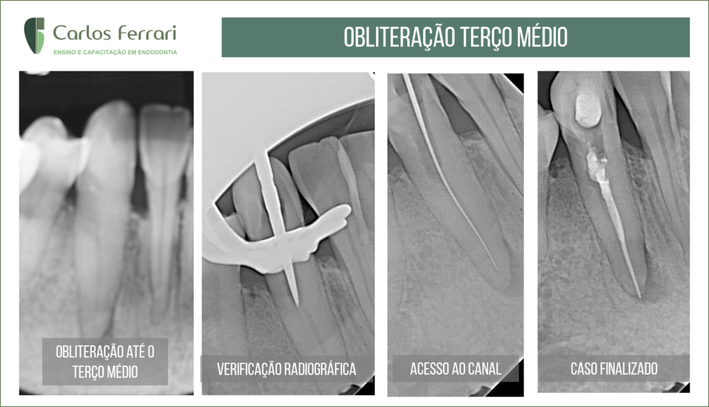
Calcificação do canal radicular. Tratamento em canino
Tratamento endodôntico com obliteração da luz do canal acessado de maneira tradicional, com pontas diamantadas finas e muita paciência
Caso realizado na turma III de especialização em endodontia na HPG Brasília pela aluna Alessandra.
Calcificação do canal radicular. In: Consolaro e Bernardini. Insight Ortodôntico • Rev. Dent. Press Ortodon. Ortop. Facial 12 (6) • Dez 2007
Uma das formas de adaptação celular a condições adversas é a metaplasia celular. A célula madura se transforma, muda seu fenótipo para um outro tipo celular igualmente maduro de célula e da mesma linhagem embrionária. A metaplasia representa uma forma eficiente de adaptação celular. Quando o indivíduo começa a fumar, as células de revestimento da traquéia e dos brônquios mudam de células epiteliais ciliadas e mucosas, cilíndricas, e grandes produtoras de muco, para células epiteliais escamosas e estratificadas, às vezes produzindo, inclusive, queratina superficial. Enfim, a metaplasia representa uma metamorfose na morfologia e função celular.
Na polpa de dentes traumatizados e que permaneceram estruturalmente hígidos, mas com lesão parcial do feixe vascular e nervoso, as células pulpares podem sofrer metaplasia para se adaptarem à hipóxia passageira e redução do metabolismo. Esta metaplasia leva os fibroblastos, os pericitos, as células indiferenciadas ou células-tronco teciduais, os pré-odontoblastos e até as células vasculares a se diferenciarem, modificarem ou se transformarem em odontoblastos, que iniciam uma produção aleatória e desorganizada de dentina reacional, com células e vasos incluídos em sua estrutura, a ponto de ser identificada até como osteodentina ou vasodentina. A osteodentina e a vasodentina são formas primitivas de dentina e encontradas como parte de dentes de animais inferiores na escala biológica evolutiva da vida animal.
Este depósito aleatório de dentina displásica, ou seja mal formada, geralmente é muito desorganizado e pouco mineralizado e pode até ter um direcionamento da periferia para o centro da polpa, mas nem sempre. Com freqüência, esta aleatoriedade de deposição de dentina displásica é absoluta e descontrolada. Após três meses, radiograficamente pode se notar apagamento dos limites pulpares normais, redução do volume pulpar, fechamento dos espaços pulpares na câmara e/ou no canal radicular . O fechamento pode se completar com o apagamento dos espaços pulpares entre 6 meses e 1 ano após o traumatismo. Este fenômeno, muito conhecido e freqüente, também é simplesmente conhecido como Obliteração Pulpar, mas deveria ser mais apropriadamente identificado como Metamorfose Cálcica da Polpa, o nome mais utilizado na literatura pertinente sobre o assunto.
Clínica e radiograficamente a presença de Metamorfose Cálcica da Polpa indica história pregressa de traumatismo dentário. Dentes traumatizados quando movimentados ortodonticamente têm mais chances de apresentarem reabsorções radiculares mais severas, pois iniciam-se mais precocemente que os demais dentes.
Calcificação do canal radicular. Tratamento em canino
https://ferrariendodontia.com.br/anatomia-endodontica/
https://www.youtube.com/watch?v=83SEaibzJuI&t=1514s