Displasia cementária periapical. Relato de caso.
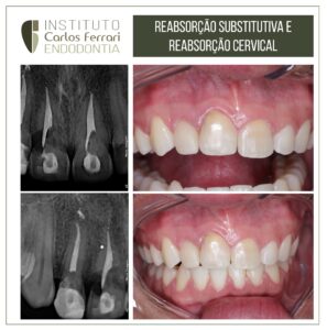
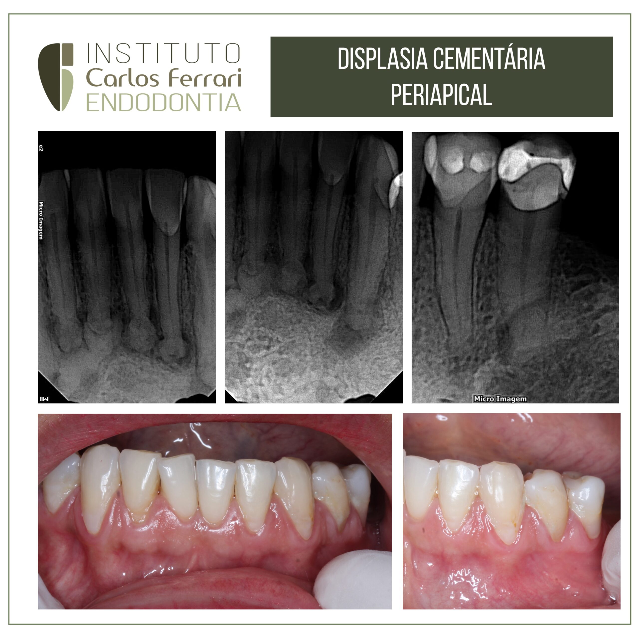
Paciente branca, 40 anos, sexo feminino, procurou a clínica por indicação de colega para tratamento endodôntico de dentes com “lesões periapicais em todos os dentes inferiores anteriores”. Nenhum sintoma foi relatado e no exame clínico, aspecto normal dos dentes e tecidos adjacentes citados. Nos exames clínicos, testes de percussão e palpação negativos e testes térmicos com resposta normal. O exame radiográfico revelou imagens radiolúcidas periapicais em todos os incisivos inferiores e também na região de 34 e 35.
Na pesquisa de antecedentes familiares, a mulher relatou a ascendência negra por avós, o que reforçou a hipótese de diagnóstico de displasia cementária periapical. A paciente foi orientada para o acompanhamento clínico e radiográfico, sem qualquer necessidade de intervenção endodôntica.
Caso conduzido pelo aluno Marcus Thurler da turma 9 de especialização em endodontia da HPG Brasília.
Displasia cementária in: Bittencourt et al. Displasia Cementária Periapical. Relato de Caso. Rev Inst Ciênc Saúde
2007; 25(3):319-21
Introdução e Revisão da literatura
A displasia cementária periapical (DCP) é um tumor odontogênico benigno de origem mesenquimal, derivado do ligamento periodontal Sua etiologia ainda permanece desconhecida embora, alguns autores, acreditam que represente uma reação incomum do osso periapical a um fator irritante local enquanto outros sugerem que o traumatismo, distúrbios hormonais, fatores sistêmicos e genéticos possam estar envolvidos no seu desenvolvimento.
.
A DCP é autolimitante, ou seja, o córtex ósseo não éexpandido e o crescimento progressivo é raro. Não há manifestações clínicas sendo, portanto, assintomáticas exceto quando localizadas próximo ao forame mentoniano, devido à compressão do nervo que pode levar a dor e parestesia . Por estas razões, esta lesão normalmente é descoberta em exames radiográficos de rotina.
Existe uma marcada predileção por mulheres (14:1) e aproximadamente 70% dos casos afetam negros. O diagnóstico nunca é feito antes dos 20 anos de idade. Nos exames imageológicos são observadas mais frequentemente em áreas correspondentes às raízes dosincisivos inferiores. Focos múltiplos são mais habituais, mas quando único, não excede 1 cm de diâmetro. Pode-se perceber ainda que a lesão está contígua ao ligamento periodontal e a lâmina dura permanece intacta.
Esta patologia possui uma evolução natural, passando por três fases de desenvolvimento: osteolítica; cementoblástica e de maturação. A 1ª fase caracteriza-sepela substituição do tecido ósseo por tecido fibroso o que determinada uma radiolucidez periapical. Na 2ªfase ocorre deposição de espículas de cemento, devidoao aumento da atividade cementoblástica, ocasionando
áreas de condensação focal opaca com áreas de rarefações. Na fase final, de maturação, há uma completa calcificação da região fornecendo uma imagem radiopaca, muitas vezes circundada por um estreito halo radiolúcido. A DCP pode levar meses ou até anos paraatingir a fase de maturação.
O tratamento é desnecessário já que a lesão estabiliza-se sem causar complicações, sendo necessário apenas à observação periódica. Sabendo-se das características da DCP, nota-se asua grande semelhança com outras patologias como as de origem endodôntica (cistos e granuloma) que sãocondições mais comumente encontradas e que se assemelham com a 1ª fase ou osteolítica.
Portanto, é essencial a realização do teste de vitalidade pulpar já queem casos de DCP a polpa encontra-se sensível aoteste, não justificando tratamento endodôntico.
Displasia cementária periapical