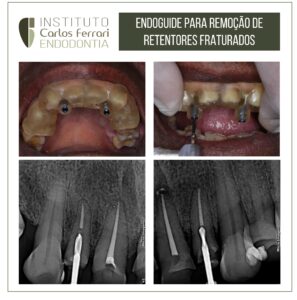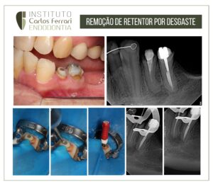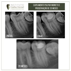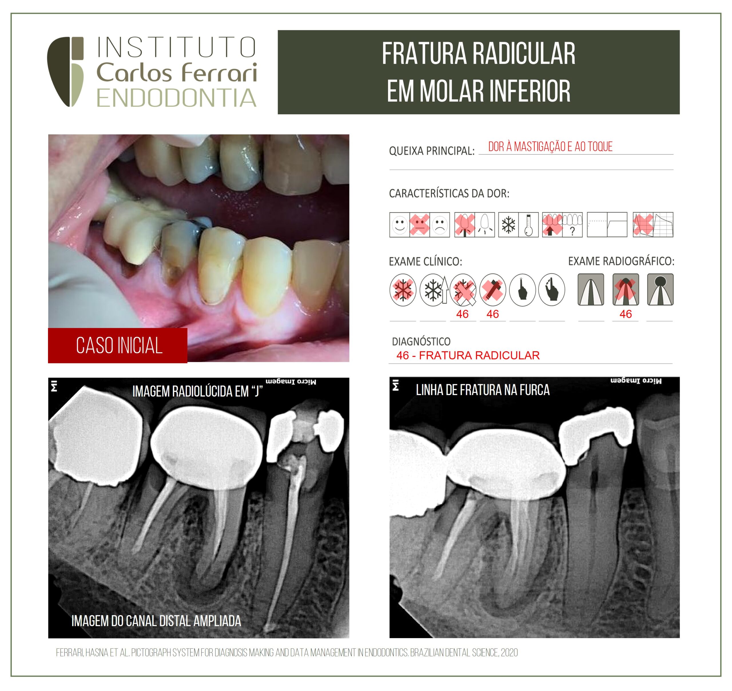
Fratura radicular em molar inferior.
Paciente indicado para tratamento endodôntico do dente 45 com finalidade restauradora, relatou dor à mastigação e ao toque no dente 46. No exame clínico observou-se coroa protética no referido dente além de recessão gengival e dor à percussão e palpação. O exame radiográfico revelou imagens compatíveis com fratura radicular: linha de fratura na furca, imagem em ¨J¨na raiz distal e imagem ampliada do canal distal.
O diagnóstico foi, portanto, de fratura radicular e o paciente encaminhado para exodontia do elemento dental.
Fratura radicular em molar inferior. In: Furtado et al. Revista Brasileira de Pesquisa em Saúde 2010; 12(2) : 61-68 |
RESUMO Um dos problemas que merece destaque na endodontia é o diagnóstico tardio das fraturas radiculares verticais. Objetivo: Alertar o cirurgião-dentista clínico-geral sobre a importância do diagnóstico precoce de fraturas radiculares verticais (FRV) prevenindo perdas dentárias futuras. Materiais e métodos: Por meio de uma pesquisa em livros didáticos, na base de dados Medline e em outros sites científicos, realizou-se uma revisão de literatura sobre o diagnóstico das FRV. Resultados: Constatou-se que os dentes tratados endodonticamente, portadores de retentores intrarradiculares inadequados e pilares de prótese fixa são os dentes mais acometidos. Os pré-molares superiores e os pacientes com idade média de 50 anos são os mais prevalentes. As causas mais comuns são devido a fatores iatrogênicos. No exame clínico, destacam-se a sondagem de bolsa periodontal, a fistulografia, a transiluminação e os testes de mobilidade e vitalidade pulpar. No exame radiográfico, pode-se observar um espessamento do ligamento periodontal, radiolucidez periapical e separação dos fragmentos da raiz. Um grande auxiliar no diagnóstico é a tomografia computadorizada, porém, em caso de dúvida, deve-se sempre lançar mão da cirurgia exploratória. Conclusão: Conclui-se que para o diagnóstico precoce das FRV, tem-se que valorizar, minuciosamente, cada uma das etapas da semiologia subjetiva e objetiva.
INTRODUÇÂO: As fraturas radiculares verticais (FRV) são um desafio para o cirurgião-dentista quanto à sua detecção precoce e conduta a ser seguida. Diagnosticar essas fraturas é essencial antes de qualquer tratamento endodôntico ou restaurador tendo
em vista que elas podem afetar drasticamente o sucesso do tratamento. São caracterizadas por uma completa ou incompleta linha de fratura que segue longitudinalmente no longo eixo do dente em direção apical.
Frequentemente,estende-se através da polpa e do periodonto.
As fraturas representam 10,9% das causas de extrações dentárias, com maior incidência em dentes tratados endodonticamente.
O diagnóstico de FRV geralmente se torna difícil pois não há sinais, sintomas e características radiográficas exatas, podendo ser confundido com um insucesso no tratamento endodôntico e até mesmo uma doença periodontal. Os sinais e sintomas mais comuns em FRV em dentes tratados endodonticamente são dor, edema, presença de fístula e uma bolsa periodontal isolada, profunda e estreita. Já as características radiográficas são representadas pelo espessamento do ligamento periodontal; perda óssea vertical, localizada e profunda; perda óssea perirradicular localizada (halo de perda óssea).
Fratura radicular em molar inferior.
https://ferrariendodontia.com.br/fratura-radicular-de-molar/