Lesão periapical sem origem endodôntica.
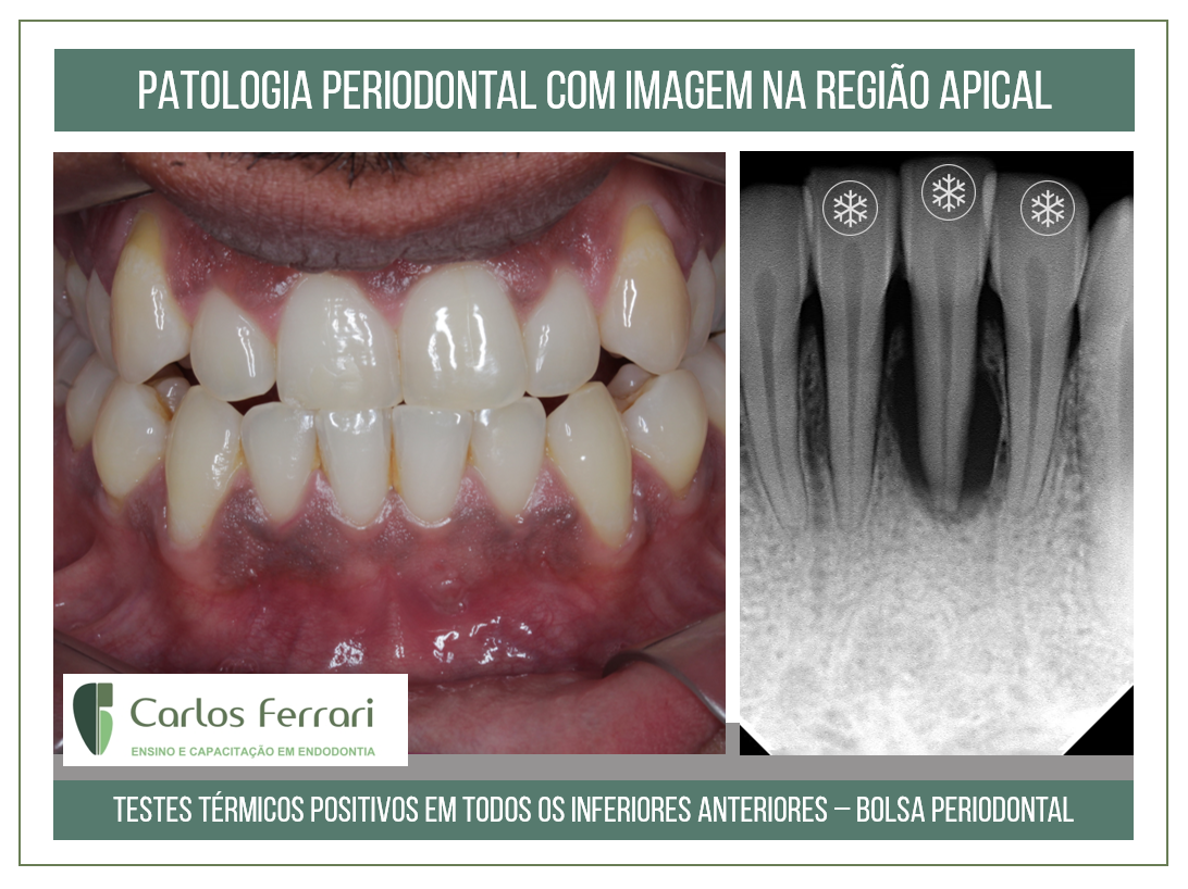
Paciente enviado para tratamento endodôntico do elemento 31 em virtude de observação de imagem radiolúcida periapical. No exame clínico, os testes não revelaram qualquer resposta diferente em relação aos dentes contíguos, positivos em relação ao teste térmico com gás e negativos à percussão. Leve dor foi relatada em relação ao teste de palpação na região radicular do dente 31. Na sondagem, foi revelada bolsa periodontal nas regiões proximais. Observa-se também presença de cálculos.
A patologia foi diagnosticada como de origem periodontal e o caso enviado ao periodontista para tratamento e orientação para possível tratamento endodôntico concomitante ao periodontal.
In: Ferrari et al. Radiologia aplicada à Endodontia, Machado, R. Endodontia. Princípios Biológicos e Técnicos. Ed. Gen, 2022.
Patologias não endodônticas com manifestações radiográficas periapicais
Patologias ou alterações periapicais não causadas por comprometimentos pulpoperirradiculares podem apresentar imagens frequentemente confundidas como tais. Um exame completo do paciente, considerando inicialmente sua queixa inicial, o histórico do caso e os resultados de um minucioso exame clínico, podem, na maioria das vezes, fornecer dados que permitam um correto diagnóstico. A ausência de sinais ou sintomas e respostas normais aos testes de vitalidade, percussão e palpação em todos os dentes adjacentes à lesão já podem constituir um substancial indicativo de sua origem não endodôntica.
A interpretação radiográfica deve ser realizada com atenção e rigor, seguindo critérios específicos e objetivos, dentre os quais destacam-se: número de lesões, localização, radiolucidez, características das bordas, estruturas internas e efeitos sobre estruturas e dentes adjacentes. Tais quesitos, quando analisados em ordem, facilitam a determinação de hipóteses diagnósticas.
As patologias e as alterações de origem não endodôntica mais comumente diagnosticadas de maneira equivocada como tal são descritas a seguir, de acordo com sua radiolucidez.
Radiopacas
•Osteosclerose idiopática: imagem única, normalmente presente na região periapical dos dentes posteroinferiores, com radiopacidade variável, bordas irregulares e ausência de cortical. Não tem etiologia conhecida, nem requer tratamento, sendo frequentemente confundida com a osteíte condensante, causada por um processo inflamatório crônico de origem endodôntica
•Tórus mandibular: formações ósseas externas localizadas na superfície lingual mandibular conseguem produzir no exame periapical imagens radiopacas com número variável e formato arredondado, frequentemente sobrepondo-se à região periapical dos dentes inferiores. O exame clínico pode confirmar a suspeita ao se observar tais formações por meio da inspeção intraoral.
Mistas
•Displasias cemento ósseas: lesões idiopáticas nas quais há substituição do tecido ósseo por tecido fibroso. Apresentam imagem mista, sendo mais radiolúcidas na primeira fase, tornando-se mistas e em seguida radiopacas após a maturação. São mais comuns em pacientes negras, do gênero feminino e em idade adulta, autolimitantes e frequentemente diagnosticadas por meio de radiografias de rotina. Contudo, por localizarem-se na região periapical, sua presença é comumente associada à realização de tratamentos endodônticos desnecessários. As displasias cemento-ósseas subdividem-se em displasia cemento-óssea periapical, mais localizada em apenas uma região (frequentemente nos dentes anteroinferiores), e displasia cemento-óssea florida, com múltiplas imagens espalhadas pela arcada.
•Outras patologias com imagens radiográficas mistas que podem mimetizar lesões de origem endodôntica são os odontomas e o cementoblastoma benigno.
Lesão periapical.