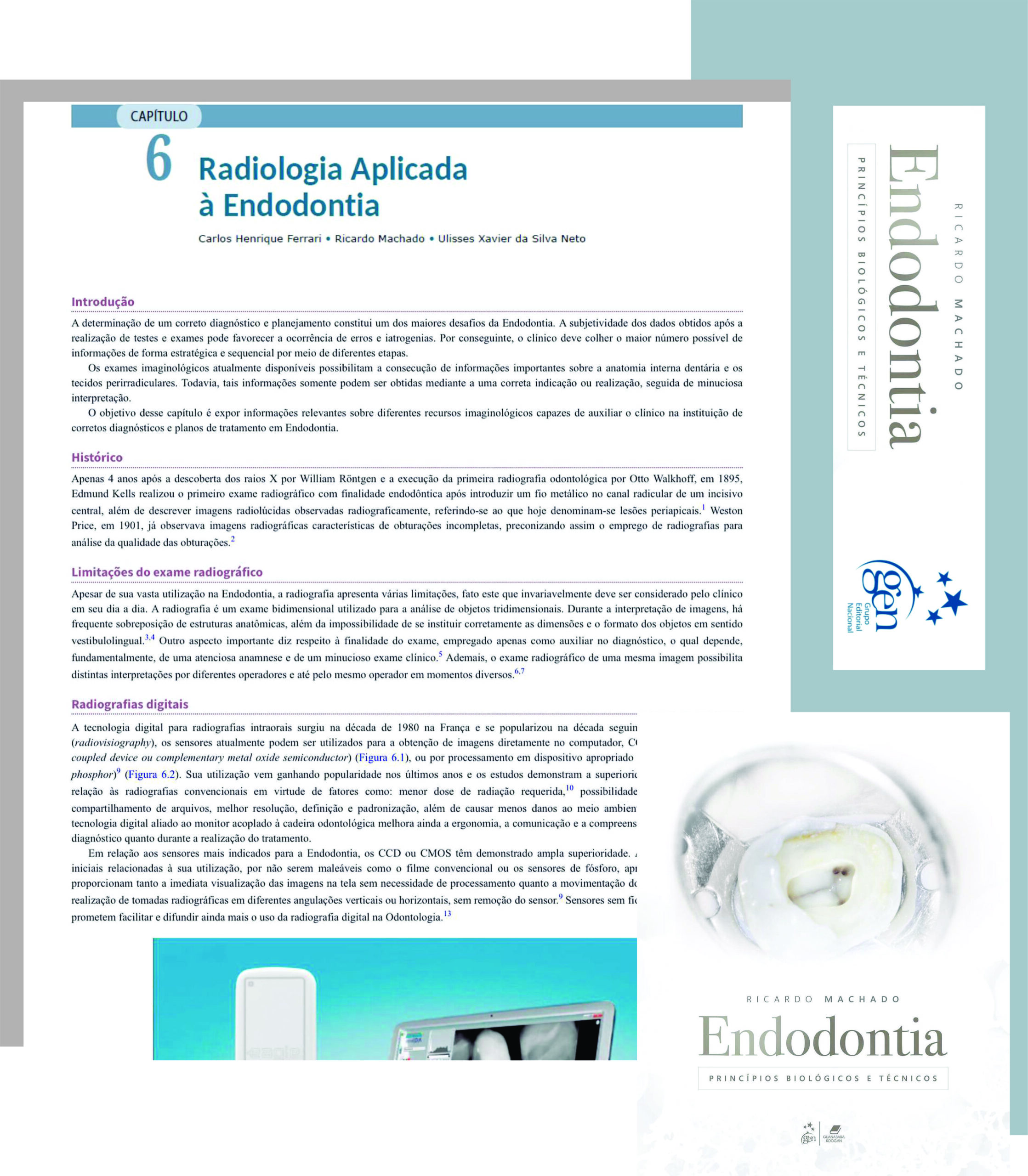
Lançamento do livro de endodontia do Prof. Ricardo Machado conta com o capítulo da nossa autoria: Radiologia Aplicada à Endodontia.
Desde os primórdios da endodontia, certas descobertas foram decisivas para sua evolução, de um simples ato empírico na ânsia da remoção da dor até os tratamentos que hoje dispomos. Um marco decisivo, senão o mais importante, foi a descoberta dos raios-x e sua consequente utilização na odontologia e no tratamento endodôntico. A radiologia, portanto, levou a endodontia ao nível de ciência como hoje conhecemos.
O exame radiográfico é o recurso que permite ao clínico o planejamento e a observação das diversas etapas do seu trabalho, bem como a avaliação final e a comunicação entre seus colegas e pacientes. Possibilita ainda a rápida avaliação dos tecidos dentais, da anatomia do canal radicular, dos tecidos ósseos adjacentes, bem como a determinação do comprimento do trabalho endodôntico e do ajuste do cone de obturação, além da avaliação final e posterior do tratamento.
Histórico
Apenas 4 anos após a descoberta dos raios-x por William Röntgen e a realização da primeira radiografia odontológica por Otto Walkhoff, em 1895, Edmund Kells realizou a primeira radiografia com finalidade endodôntica, ao realizar uma tomada após introduzir um fio metálico em um canal radicular de um incisivo central. Também descreveu imagens radiográficas radiolúcidas, características de lesões no periápice. Já Weston Price, em 1901 já observava a imagem radiográfica característica de obturação incompleta, preconizando pela primeira vez a utilização da radiografia para verificação da qualidade da obturação endodôntica 2.
Limitações do exame radiográfico
Apesar de sua vasta utilização na endodontia, a radiografia possui algumas limitações. Tal fato serve como alerta constante para o clínico na interpretação das imagens.
A radiografia é um exame bidimensional utilizado para a observação de corpos que possuem três dimensões. Tal fato tem que ser constantemente levado em consideração pelo observador, já que a interpretação de tais imagens tem que levar em conta as sobreposições de diferentes estruturas anatômicas sobre as áreas de interesse. Além disso, não é possível a correta estimativa das dimensões e formato das imagens em sentido vestibular/lingual 3, 4. Outro aspecto diz respeito sobre a finalidade do exame, utilizada apenas como auxiliar no diagnóstico, ficando na dependência de outros exames complementares e principalmente do histórico do caso e exame clínico. Além disso, o exame radiográfico permite diferentes interpretações por observadores e até mesmo pelo mesmo observador em momentos distintos.
https://ferrariendodontia.com.br/curso-online-radiologia-endodontia/