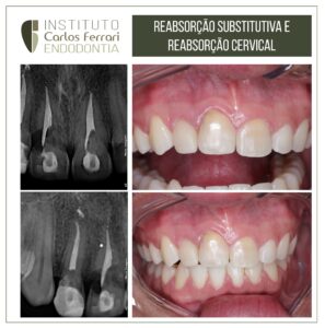
Retentores intrarradiculares. Retratamento em dente com retentor de fibra.
Dentes com retentores de fibra costumam ser um desafio ao retratamento.
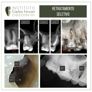
Paciente cirurgiã dentista, procurou a clínica pra retratamento de dente 11 com fístula, tratado há 3 anos e com um pino de fibra no interior do canal. Com a utilização de microscópio e pontas de ultrassom, o retentor foi ultrapassado e desgastado para o retratamento endodôntico do canal.
In: Machado, Ricardo. Endodontia: Princípios Biológicos e Técnicos. Disponível em: Grupo GEN, Grupo GEN, 2022.
Retentores intrarradiculares estéticos*
Retentores intrarradiculares cerâmicos também são considerados estéticos. Porém, a seguir, o termo “estéticos” refere-se a pinos de fibra.
Pinos estéticos têm sido cada vez mais utilizados, sobretudo por apresentarem comportamento biomecânico mais favorável e estética superior, além de demandarem menor tempo clínico para a confecção e instalação.51
Diante da necessidade de remoção, as características e o método de cimentação são determinantes para o sucesso do procedimento, visto que os cimentos resinosos são mais dificilmente removidos em razão de sua natureza viscoelástica e resiliência, que atenuam as vibrações ultrassônicas.
Os materiais mais comumente utilizados para a fabricação de pinos estéticos são as fibras de vidro, quartzo e carbono com quartzo. Por serem escuros, os pinos de carbono podem receber um revestimento com fibra de quartzo ou vidro, o que melhora a estética, sem comprometer suas propriedades positivas. Os pinos de fibra de vidro ou quartzo são mais baratos que os de fibra de carbono, e apresentam certa capacidade de transmissão de luz. Porém, para a sua cimentação, são necessários um adesivo e um cimento de presa química ou dual, pois a luz transmitida não alcança o terço apical.52
Os protocolos para a cimentação de pinos estéticos pautam-se nos preceitos da Odontologia Adesiva, em que uma camada híbrida é formada no interior do canal para que os pinos sejam adesivamente fixados à dentina intrarradicular. Desse modo, sua remoção somente pode ser realizada por desgaste.
O desgaste de pinos estéticos cimentados é considerado um procedimento de alta complexidade, em razão do eminente risco de fragilização e perfuração radicular, sobretudo, a partir do uso de brocas de alta rotação. Destarte, recomenda-se que esse procedimento seja realizado com pontas ultrassônicas e “à luz da microscopia operatória”.
Considerações iniciais para a remoção de pinos estéticos
Por meio da radiografia inicial, deve-se identificar o tipo e as características do retentor (serrilhado, cilíndrico, liso, cônico, com ou sem fio-guia etc.). Geralmente, as cores do pino e da dentina são distintas, o que contribui para que somente o pino seja desgastado, preservando as paredes radiculares. A presença de um fio-guia (fio metálico localizado no centro do pino em todo o seu comprimento) favorece à remoção sem desgastes excessivos nas paredes do canal.
Outro fator importante que deve ser ponderado para a remoção de pinos estéticos é a extensão. Quanto maior o comprimento do pino, mais difícil é a remoção em virtude do estreitamento das paredes radiculares no sentido cervicoapical. Nesse contexto, a inclinação do dente na cavidade oral também deve ser considerada.
Seleção dos insertos ultrassônicos para o desgaste
Os insertos mais utilizados para o desgaste e a remoção de pinos estéticos são os diamantados. Contudo, os lisos também podem ser empregados, embora apresentem menor capacidade de corte, sendo, portanto, mais seguros. Os insertos devem ser longos e finos para desgastar todo o retentor, preservando as paredes do canal. A irrigação durante a ativação ultrassônica compromete a visualização, razão pela qual não deve ser utilizada. Assim, recomenda-se a ativação ultrassônica por 20 segundos, intercalada com a irrigação convencional para a limpeza do canal e o resfriamento radicular.
Remoção de pinos estéticos
1.Radiografia inicial.
2.Análise e identificação dos seguintes elementos: pino, cimento, dente e resina.
3.Desgaste do núcleo e acesso ao retentor.
4.Início do desgaste do pino com uma ponta diamantada esférica de haste longa e de tamanho compatível, considerando a inclinação do dente e do pino.
5.Continuação do desgaste do pino com insertos ultrassônicos esféricos diamantados ou lisos (E9, E5, E4D e E2D – Helse Ultrasonic) com potência de 20 a 40%. Especial atenção deve ser dada à diferença de cor entre a dentina e o pino para evitar desgastes nas paredes radiculares.
6.O diâmetro das pontas de ultrassom deve ser compatível com o do canal em cada terço radicular. A realização de radiografias transoperatórias é fortemente recomendada.
7.Finalizado o desgaste do retentor, procede-se à limpeza do cimento remanescente nas paredes radiculares com um inserto ultrassônico liso na potência mínima.
8.Radiografia final.
Retentor intrarradicular