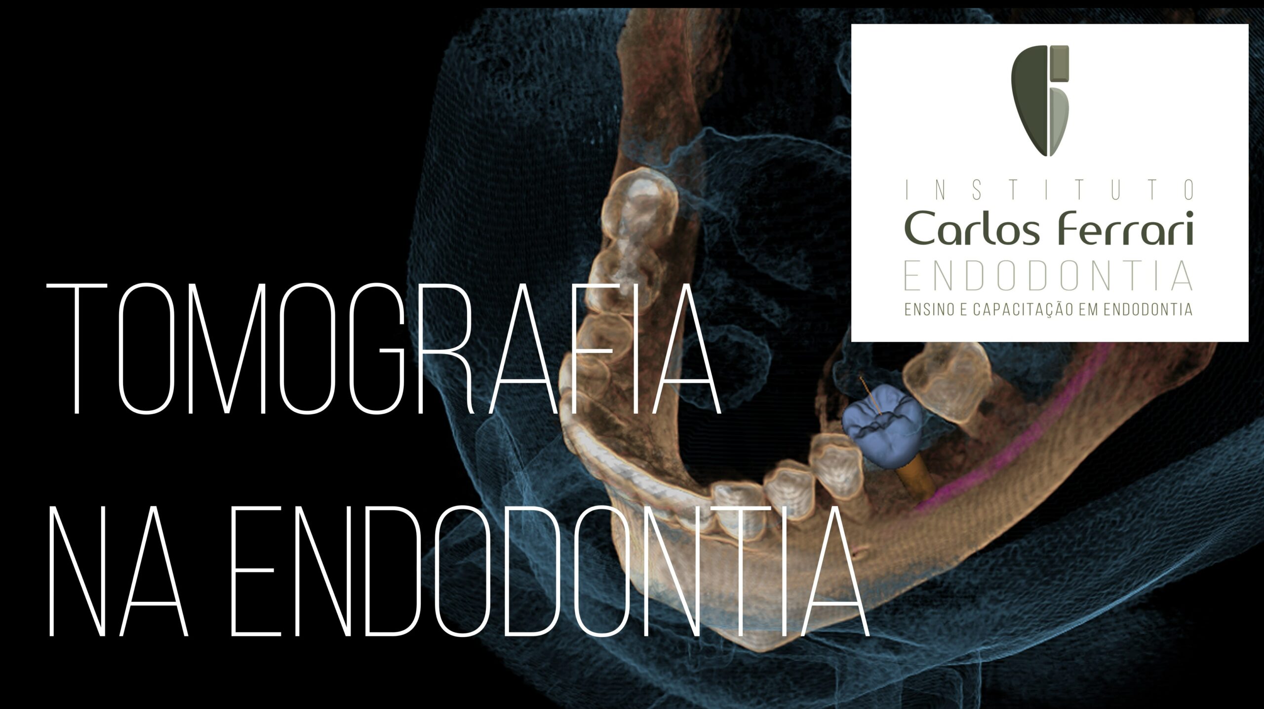
Tomografia na endodontia. 1a. parte. Aula Online no canal Carlos Ferrari Endodontia no youtube.
In: Carlos Ferrari et al. Cap. Radiologia na Endodontia. Ricardo Machado. Endodontia. Princípios Técnicose Biológicos. Ed. Gen, 2022:
Tomografia Computadorizada em Endodontia
Apesar da aplicabilidade do exame radiográfico nas diferentes fases do tratamento ou retratamento endodôntico (convencional e cirúrgico), deve-se considerar suas limitações. O advento da tomografia computadorizada proporcionou importantes avanços às diferentes especialidades odontológicas, sobretudo, devido à tridimensionalidade das imagens obtidas. Porém, o alto custo, o difícil acesso e a baixa resolução das imagens ainda representavam um impedimento quanto à sua utilização na rotina clínica.
O desenvolvimento da tomografia computadorizada de feixe cônico (TCFC) proporcionou maior acessibilidade ao exame em razão da diminuição dos custos anteriormente praticados, menores doses de radiação e maior qualidade das imagens. Na Endodontia, sua utilização passou a se popularizar com o surgimento de aparelhos com resolução ainda mais acurada e imagens com melhores detalhamento e nitidez. Portanto, há uma tendência de que TCFC seja utilizada para todos os casos, desde o diagnóstico até a proservação.
A TCFC apresenta inúmeras vantagens, como visualização tridimensional, ausência de sobreposições, possibilidade de observação em múltiplos planos e fidelidade dimensional, além de facilitar a comunicação entre o profissional e o paciente e entre profissionais.
Contudo, o exame apresenta desvantagens relevantes, como o custo, ainda relativamente elevado em comparação com o do exame radiográfico, e a produção de artefatos de imagem (interferências causadas pela presença de metal). Essas desvantagens tendem a diminuir com o tempo, com a popularização dos exames e o desenvolvimento de novas tecnologias e softwares.
Conceitos gerais
Voxel e FOV
A menor unidade da imagem tridimensional e, por consequência, utilizada na tomografia denomina-se voxel, a qual dispõe de três dimensões e formato cúbico. Quanto menor o voxel de uma imagem, melhor a sua definição. Portanto, para a Endodontia, o ideal é que as imagens sejam formadas pelas menores unidades de voxel. Já o FOV (field of view) refere-se ao tamanho da área que será exposta e fará parte do exame. Para a Endodontia, o FOV pode ter a partir de cerca de 4 × 4 cm, a fim de englobar o dente a ser estudado e a região adjacente.
Aquisição de imagens
Na TCFC, há um feixe de raios X em formato cônico que, ao girar ao redor do paciente, produz uma série de imagens que serão agrupadas pelo software, dando posteriormente, origem a uma imagem tridimensional da área estudada. Depois, essa imagem é processada para constituir os cortes e as reconstruções. As imagens obtidas são classificadas em hipodensas, isodensas e hiperdensas, de acordo com a densidade, semelhantemente às radiolúcidas, neutras e radiopacas, utilizadas nas radiografias.
Cortes e reconstruções
Após a aquisição das imagens com vistas à obtenção de uma única imagem tridimensional, pode-se investigar a área/dente em diferentes planos, utilizando filtros e outros recursos gráficos. Os cortes básicos são o sagital, o coronal e o axial, sendo o último de maior relevância para o diagnóstico endodôntico. Além deles, pode-se realizar cortes transversais, em quaisquer plano e direção.
Por meio do software de tratamento de imagens, é possível também fazer a reconstrução panorâmica, que pode ter a espessura e a área de captura da imagem personalizada. Uma reconstrução panorâmica completa (arcadas superior e inferior) é semelhante a uma radiografia panorâmica normal, porém sem sobreposições.
Outra reconstrução importante é a 3D, na qual se produz um modelo tridimensional da área capturada, com inúmeras possibilidades de tratamento, colorização, mudança de densidade e corte. Além disso, os modelos 3D podem ser impressos, para diagnósticos e planejamentos cirúrgicos.
Em resumo, as imagens tomográficas mais utilizadas em Endodontia são as proporcionadas a partir de cortes axiais e transversais, além das reconstruções em 3D.
Aplicações da tomografia computadorizada de feixe cônico em Endodontia
A TCFC pode ser utilizada em todas as fases do tratamento ou retratamento endodôntico, desde o diagnóstico até a proservação. Um exame tomográfico é capaz de fornecer um número significativamente maior de informações que uma radiografia periapical. Portanto, justifica-se o fato de que em 30 a 60% dos casos haja mudança de diagnóstico ou planejamento quando um profissional avalia uma radiografia periapical e uma TCFC do mesmo dente ou área.
A TCFC é útil para a análise tridimensional da morfologia dental, revelando com muito mais acurácia as dimensões da câmara pulpar, a quantidade e as características dos canais radiculares . Em retratamentos, é de suma importância na detecção de canais não tratados, desvios, perfurações etc.
A TCFC também pode ser utilizada para a detecção de trincas ou fraturas radiculares. Porém, nem sempre se observa a linha que confirma esta hipótese, tornando-se necessária a prospecção clínica e/ou cirúrgica. Nesses casos, um grande obstáculo para o diagnóstico reside na ocorrência de artefatos de imagem, produzindo linhas hipodensas e hiperdensas, mais frequentemente observadas em dentes portadores de retentores intrarradiculares metálicos
Em casos de reabsorções radiculares, a TCFC compreende um meio muito mais eficaz que o exame radiográfico para o seu diagnóstico. Além disso, somente um exame tridimensional pode identificar a ocorrência de comunicações entre uma reabsorção e o canal radicular e entre este e os tecidos periodontais (reabsorção externa e interna, respectivamente).
A TCFC apresenta maior acurácia na identificação de imagens hipodensas em comparação com o exame radiográfico42. Portanto, é um exame mais confiável na detecção de lesões periapicais em estágio inicial, na instituição de diagnósticos diferenciais entre lesões de origem endodôntica e não endodôntica, no planejamento cirúrgico e na proservação. A TCFC revela imagens no periápice mais precocemente, em estágios ainda não observáveis na radiografia periapical e com maior possibilidade de detecção. Em casos de imagens de maior extensão, não há consenso na literatura de que a TCFC possa constituir um método confiável na diferenciação entre cistos e granulomas periapicais.
Quando as imagens hipodensas se localizam em dentes posterossuperiores, a TCFC pode ser fundamental no diagnóstico diferencial da sinusite odontogênica, ao revelar a comunicação das lesões com o seio maxilar, o rompimento de corticais e o espessamento das mucosas adjacentes.36 Essa condição é comum e pouco diagnosticada tanto por cirurgiões-dentistas quanto por médicos.
A TCFC também pode ser utilizada para a análise da proximidade dos ápices radiculares com estruturas anatômicas nobres, como o seio maxilar e o canal mandibular. Além disso, permite a observação prévia de fenestrações apicais, a qual também pode ser útil para o diagnóstico de dores pós-operatórias de longa duração, sem causa aparente.
Em traumatismos dentais, a TCFC é útil para revelar fraturas de coroa e/ou radiculares e ósseas, bem como para identificar espaços vazios no interior do alvéolo. O exame tomográfico demonstrou ser sensivelmente mais eficiente que as radiografias panorâmicas na identificação de fraturas ósseas e alveolares e que as radiografias periapicais digitais na detecção de fraturas radiculares horizontais, inclusive com diferentes angulações.
Em casos de dentes com câmara pulpar e canal radicular atrésicos por mineralização – situação comum em dentes anteriores traumatizados – a TCFC, aliada à tecnologia de prototipagem, pode ser utilizada por meio de guias confeccionados em impressoras 3D, para nortear o acesso de uma broca cirúrgica à região periapical. Tal tratamento denomina-se acesso endodôntico guiado. Para mais detalhes, recomenda-se a leitura do Capítulo, Endodontia Guiada por CAD/CAM (Endoguide 3D).
O exame tomográfico também pode ser utilizado para o diagnóstico de corpos estranhos, instrumentos fraturados, perfurações, sobreobturação e sobre-extensão.
tomografia na endodontia
