Ápice radicular e seu posicionamento em tomografias.. Trabalho publicado.
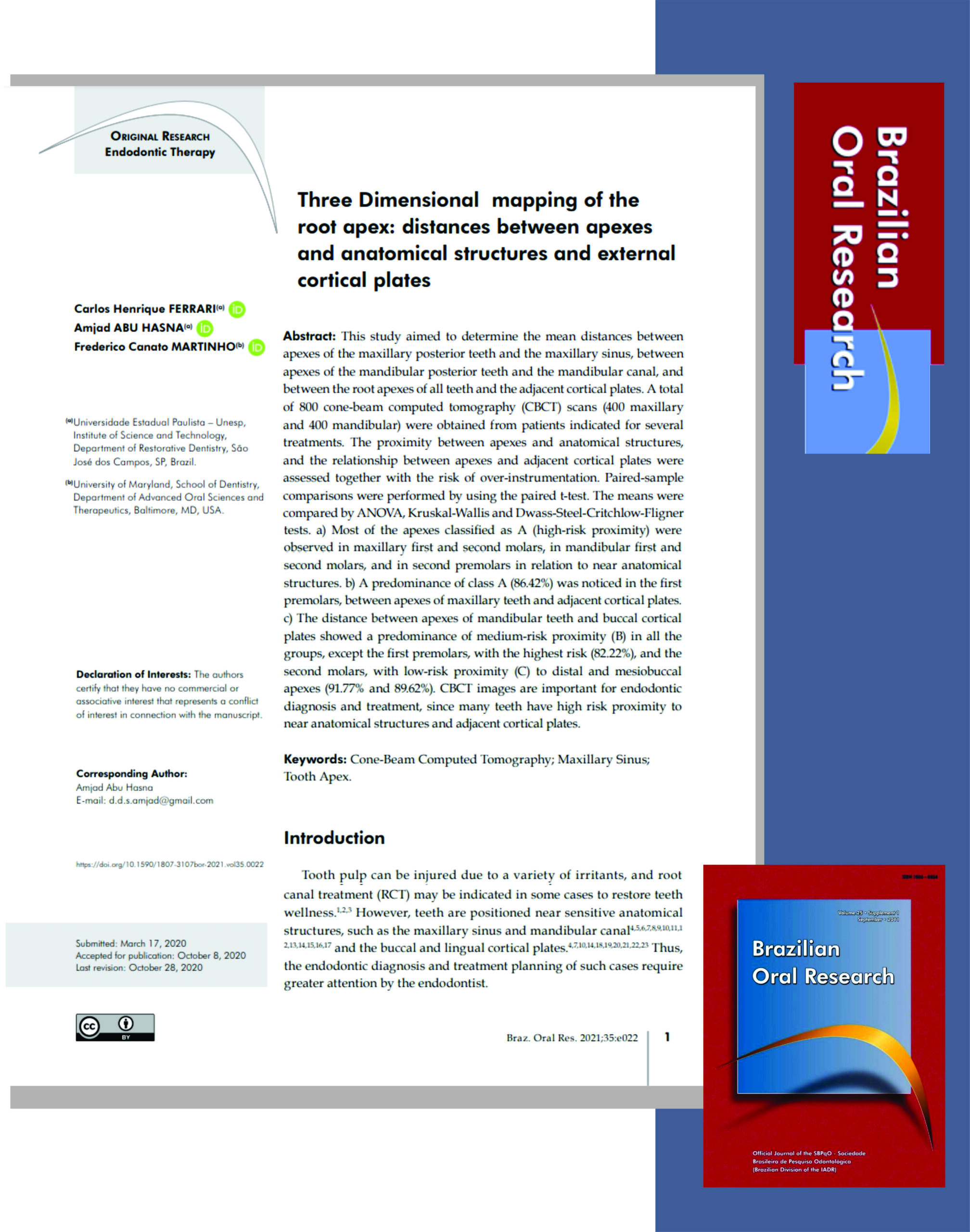
Three Dimensional mapping of the root apex: distances between apexes and anatomical structures and external cortical plates. Trabalho publicado na Revista Brazilian Oral Research, com os colegas Amjad Abu Hasna e Frederico Canato Marinho.
Assuntos conexos: ápice radicular tomografia cone beam na endodontia.
Trecho traduzido:
SUMÁRIO
O conhecimento do posicionamento dos ápices radiculares em relação às estruturas anatômicas adjacentes e às tábuas ósseas é de importância para o clínico, para um melhor planejamento do tratamento endodôntico, e consequente maior segurança nos procedimentos. Nosso trabalho teve por objetivo mapear a posição dos ápices da arcada dentária em relação às estruturas anatômicas seio maxilar e canal mandibular e em relação e às tábuas ósseas vestibular e lingual, por meio de estudo de tomografias computadorizadas de feixe cônico, e analisar os riscos da sobreinstrumentação endodôntica em relação a esses aspectos. Os resultados mostram uma porcentagem significativa de ápices em íntima relação com as estruturas citadas, alertam para os riscos de tratamentos endodônticos invasivos dada a proximidade dos ápices de determinados grupos dentais com estruturas relevantes e recomendam a utilização da tecnologia da tomografia computadorizada para o planejamento endodôntico em casos de dúvida quanto ao posicionamento apical.
Os dentes podem apresentar diferentes posições na arcada dentária, que podem levar, consequentemente, a um posicionamento dos seus ápices radiculares em regiões que requeiram maior atenção por parte do clínico, tanto no diagnóstico quanto no planejamento e execução do tratamento endodôntico. Essa proximidade se dá tanto em relação a estruturas anatômicas relevantes, como o seio maxilar e o canal mandibular quanto em relação às corticais ósseas externas, vestibular ou lingual, e, quando presente em dentes com indicação do tratamento endodôntico, representa risco de complicações e acidentes, amplamente relatados na literatura, por lesão física, química e biológica, parestesia relacionada à lesão física ou química do nervo alveolar inferior, por medicação intracanal, material obturador ou por instrumentos endodônticos, lesão química do seio maxilar, tecidos periapicais e musculares por extravasamento de substâncias químicas e a disseminação de infecções para tecidos por microorganismos oriundos do canal radicular. Tais eventos desde eventos de pequena gravidade e resolução espontânea, até acidentes de maior gravidade e resolução cirúrgica.
Além disso, os tratamentos endodônticos contemporâneos são em grande parte realizados além dos limites do canal radicular, por meio de manobras conhecidas como patência apical ou alargamento foraminal. Tais tratamentos, por seu potencial invasivo aos tecidos periapicais, mostram-se capazes de potencializar os riscos citados.
Para o exame dos ápices dentários, o recurso mais acessível é o exame radiográfico, que não oferece uma estimativa adequada da posição anatômica apical. O recurso mais adequado para tal é a tomografia computadorizada por feixe cônico (TCFC), por permitir a observação tridimensional da anatomia dentária e sua relação com a totalidade da região anatômica adjacente, eliminando sobreposições anatômicas e distorções na imagem. Porém, a TCFC ainda é um recurso pouco utilizado na clínica e a indicação desse exame como recurso de rotina no diagnóstico endodôntico não é definitivamente estabelecida pela literatura.
Ápice radicular


