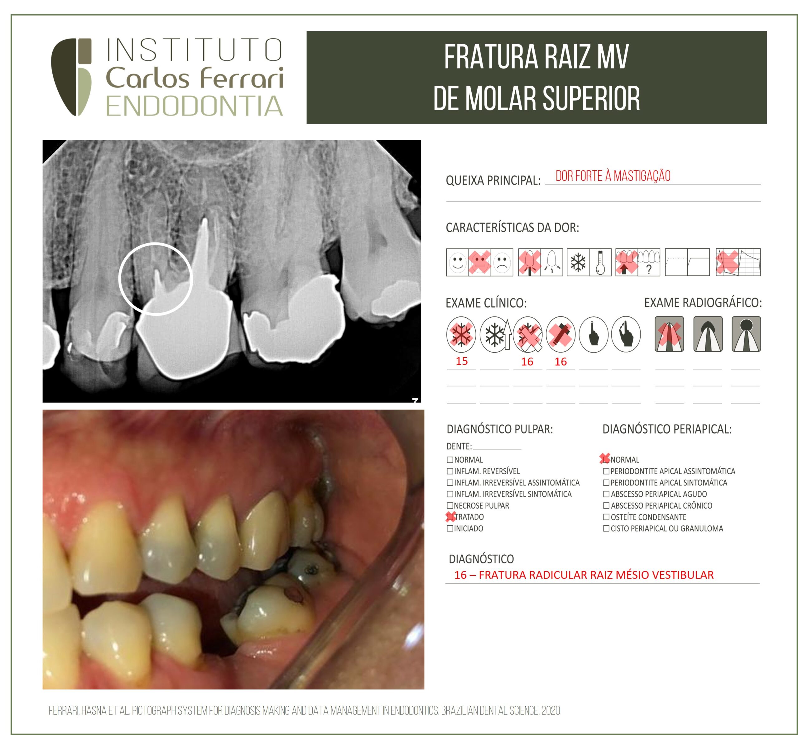
Fratura radicular de molar. Relato de caso.
Paciente veio à clínica queixando-se de dor localizada no dente 16, que o incomodava há mais de um ano. Ao exame clínico, apenas leve dor à palpação digital na região radicular MV. Observa-se oclusão do referido dente em excesso de carga em relação ao antagonista, inclinado. O exame radiográfico revelou linha de fratura na raiz MV.
Um preparo e núcleo nessas dimensões favorecem a trinca na raiz em longo prazo por concentrar forças que seriam em áreas delicadas da raiz.
Fratura radicular de molar. In: Lima et al. Research, Society and Development, v. 10, n. 12, e59101219988, 2021
Introdução
As Fraturas Radiculares Horizontais (FRH) resultam, geralmente, de um impacto frontal decorrente de traumas por objeto duro ou luta. Abrangendo de 1,2 -7% de todas as lesões traumáticas, essas, ocorrem em dentes permanentes (Andreasen, Andreasen & Tsilingaridis, 2018; Andreasen, 1979) e afetam, normalmente, um único elemento, conforme elucidado no trabalho de Andreasen, Andreasen e Cvek (2007).
Os incisivos centrais superiores são acometidos com mais frequência, seguido dos incisivos laterais superiores e apenas em 5% dos casos em incisivos inferiores (Andreasen, Andreasen & Bayer, 1989).
Tradicionalmente, as fraturas radiculares horizontais eram diagnosticadas apenas por imagens radiográficas e classificadas de acordo com a posição da linha de fratura, podendo estar localizada no terço apical, médio ou cervical (Caliskan & Pehlivan, 1996; Wolner-Hansen, 2010). Pode-se, ainda, ser definida como oblíqua, quando envolve o terço cervical e médio, ou transversal, quando se restringe apenas a um terço do dente (Cvek, Andreasen & Borum, 2001).
Diante da necessidade de um melhor diagnóstico por imagem, as técnicas radiográficas alcançaram grandes desenvolvimentos, sendo atualmente representadas por imagens tridimensionais (Costa et al., 2014).
A Tomografia Computadorizada do tipo Cone-Beam (TCCB) tem sido utilizada com sucesso no diagnóstico e
prognóstico de imagem das fraturas radiculares e se mostra superior a outros métodos radiográficos (Fagundes, Mendonça, Albuquerque & Jacinto, 2014).
Foram realizados estudos comparando a tomografia computadorizada (TC) e radiografias para o diagnóstico de fraturas radiculares e foi demonstrado que o nível da linha de fratura em radiografias pode variar significativamente em relação
à imagem tridimensional. Sendo assim, quando a exata posição da fratura horizontal não puder ser determinada, o diagnóstico
radiográfico deve ser complementado com a imagem tridimensional, o que poderá afetar o planejamento do tratamento (Bornstein, Wolner- Hanssen, Sendi & Arx, 2009; Bernardes et.al, 2009).
Com isso, houve uma melhora significativa no diagnóstico de fraturas radiculares horizontais, com relação a presença e ausência dessas, a exata localização, extensão e direção da linha de fratura (Lenzi & Trope, 2012; Costa et al., 2011).
Fraturas radiculares geralmente apresentam um bom prognóstico, sendo caracterizada por uma comunicação entre os tecidos pulpares e periodontais, formando, assim, uma linha de fratura. Esse processo promove o reestabelecimento de uma circulação colateral e drena o edema causado pelo trauma, assim como ocorre a diminuição da tensão nos vasos pulpares
(Mata, Gross & Koren, 1985). A vitalidade pulpar normalmente é mantida após fratura radicular, ocasionando cicatrização
espontânea de 70 a 80% dos casos (Andreasen, & Andersson, 2007; Camp, 2000).
O reparo das fraturas radiculares horizontais envolve a união entre os segmentos por tecidos duros, calcificados (ocorre raramente), interposição de tecido conjuntivo (ocorre comumente), interposição de tecido ósseo e conjuntivo ou interposição de tecido de granulação (Neeraj, Kundabala & Acharaya, 2011).
A separação entre os fragmentos é uma variável importante na cicatrização da fratura (Ozbek, Serper &Semra, 2003).
Se o deslocamento do fragmento coronário não for severo, ocorrerá um dano mínimo a polpa e ao periodonto. Na presença de
um espaço entre os fragmentos, o reposicionamento dos mesmos aumenta a frequência de cicatrização, particularmente em
dentes com formação radicular completa. Em dentes com formação radicular incompleta, uma regeneração tecidual irá ocorrer,
mesmo se o espaço da fratura for persistente, pois há uma excelente capacidade pulpar de revascularização (Andrade, Campos
Sobrinho, Andrade & Matos, 2008).
A ocorrência da cicatrização continua a ser a mesma, independentemente do local da fratura em relação ao sulco
gengival (Cvek, Mejare & Andreasen, 2002). No entanto, no caso de uma invasão bacteriana proveniente do sulco gengival, a
fratura na região cervical está mais propensa à contaminação devido a sua proximidade com o sulco. O reparo da fratura por tecido duro é menos frequente no terço cervical que em outras partes da raiz, provavelmente
devido a maior mobilidade do fragmento coronário (Cvek et al., 2001; Cvek et al., 2002). Além disso, o prognóstico é
significativamente melhor nas fraturas oblíquas localizadas na cervical que as fraturas transversais localizadas nesta mesma
região (Cvek et al., 2002), considerando que nas fraturas oblíquas, o envolvimento do terço médio ajuda a estabilizar o
fragmento coronário (Fagundes et al., 2014).
Com relação ao tratamento, é proposto que haja uma redução da fratura, que consiste no alinhamento e justaposição
entre os fragmentos, com estabilização do fragmento coronário com auxílio de esplintagem. Esse procedimento visa permitir a
formação de uma ponte de tecido calcificado entre os fragmentos (Cvek et al., 2001). É recomendável a esplintagem do dente
utilizando de uma fixação flexível por 4 semanas, se a fratura for localizada no terço médio ou apical. Se a fratura estiver
localizada cervicalmente, a esplintagem permanece por um período de tempo maior (até 4 meses) (Bourguignon et al., 2020;
Cvek et al., 2001; Diangelis, Andreasen & Ebeleseder, 2012).
Torna-se de grande relevância o embasamento teórico frente ao caso clínico tratado nesse trabalho, para que o
acréscimo de conhecimento possa auxiliar a prática clínica diária, frente a situações de urgência odontológica de pacientes
traumatizados, em específico da FRH em dentes permanentes. Como resultado da teoria em concomitância com a prática,
obteremos diagnósticos mais precisos, tratamentos adequados e prognóstico o mais positivo possível. Sendo assim, é de suma
importância a compreensão acerca dos mecanismos que levam a este tipo de fratura, às respostas teciduais, bem como o
manejo dos diferentes tipos de fratura e os respectivos prognósticos (Abott, 2019).
Fratura radicular de molar.