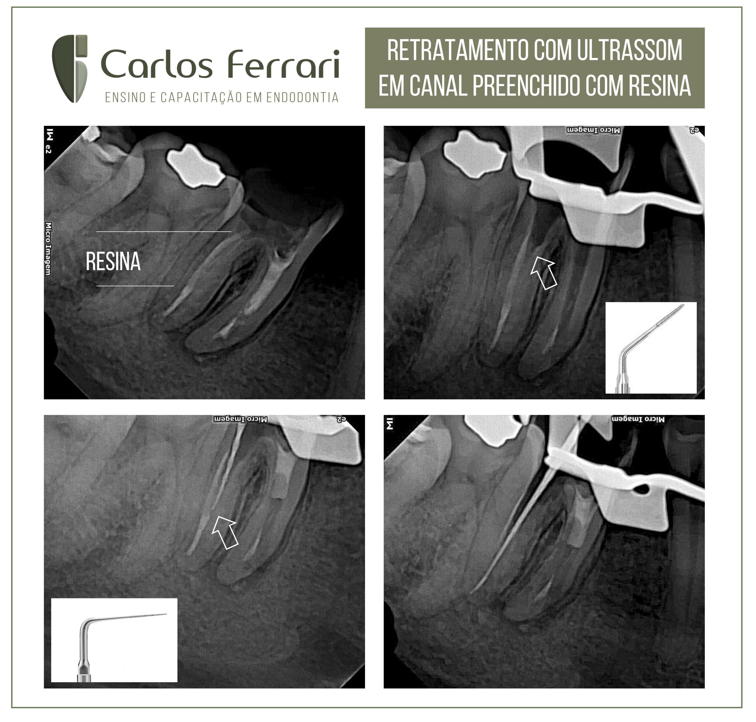
Retratamento com ultrassom.
Caso de retratamento com ukltrassom em que a presença de resina no interior do canal radicular dificultou o seu esvaziamento. Paciente procurou a clínica de especialização em endodontia, com quadro de periodontite apical sintomática e canal tratado. Durante o esvaziamento do canal, houve grande dificuldade no terço cervical do canal distal, pela presença de material endurecido, compatível com resina. Aplicou-se então força moderada com pontas de ultrassom diamantadas, maiores no início e de menor calibre no terço apical, até que se tivesse êxito na ultrapassagem com o instrumento até forame.
Caso conduzido pela aluna Camila Soares do curso de especialização em endodontia da HPG Brasília.
Ultrassom na endodontia
O uso dos aparelhos de ultrassom na odontologia têm se difundido muito nos últimos tempos. Na endodontia, certamente são múltiplos os seus benefícios na tentativa de otimizar, simplificar e aumentar a eficiência do preparo químico-mecânico do sistema de canais radiculares.
Os efeitos biológicos do ultrassom podem causar alterações teciduais, celulares e estruturais locais. Esses efeitos são provenientes da geração de calor, produção de correntes acústicas além da cavitação produzida durante o seu uso.
Tipos de Ultrassom
Antes de mais nada, é importante falarmos sobre os diferentes tipos de ultrassom. A produção da energia ultrassônica para a área odontológica pode ser feita por diferentes sistemas de transdutores: o magnetostritivo e o piezoelétrico.
O sistema magnetostritivo foi o primeiro sistema de geração de vibração ultrassônica utilizado em odontologia. Neste sistema, a vibração é gerada através da incidência de uma corrente elétrica em placas metálicas justapostas. Desse modo, a energia causa a vibração das placas e é então transferida para um inserto ou ponta de ultrassom que, por sua vez, irá se movimentar. Por isso, este sistema gera muito calor durante seu uso.
Na busca evolutiva por um sistema mais adequado para o uso odontológico, a tecnologia ultrassônica passou a utilizar o chamado sistema piezolétrico. Neste sistema a vibração é resultante da aplicação de um campo elétrico sobre cristais de quartzo ou a turmalina. Estes cristais quando expostos a um campo elétrico alternado sofrem variações em sua espessura, se expandindo e contraindo.
Dessa forma, estes movimentos de expansão e contração geram ondas sonoras ultrassônicas que podem ser aproveitadas para a produção de vibração. Assim sendo, cada transdutor possui uma frequência de ressonância diferente.
Quanto menor a espessura do cristal de quartzo, maior a sua frequência de vibração. A maioria dos aparelhos ultrassônicos piezoelétricos disponíveis no mercado trabalham em frequência superiores a 30.000Hz. Em suma, entende-se por frequência ultrassônica o número de movimentos realizados em um determinado intervalo. A frequência não deve ser confundida com potência. Assim sendo, a potência ultrassônica está relacionada com a amplitude do movimento produzido.
>>>Leia mais: Motor piezoelétrico: o aliado que todo cirurgião precisa ter
Aparelhos piezoelétricos
Nos aparelhos de ultrassom piezelétricos não cirúrgicos -convencionais- (figura 1), disponíveis no mercado atual, é possível a realização do ajuste da potência, porém, não da frequência de vibração. Portanto, com o aumento da potência, a amplitude da oscilação tende a aumentar também.
Estes aparelhos são mais precisos no corte do que os magnetostritivos, ao passo que gera menos calor sendo biologicamente mais compatível. Além disso, podem ser usados em cirurgias paraendodônticas microscópicas, visto que permitem cortes de tecidos duros, como dentina e osso, sem danificar os tecidos moles.
Utilização do Ultrassom na endodontia
A frequente ocorrência de problemas na clínica como fraturas de instrumentos, perfurações radiculares, formações de zips, desgastes inadequados e não uniformes dentro dos canais radiculares, permitem que possamos utilizar os insertos ultrassônicos justamente para solucionar estes problemas.
O desenvolvimento de novos insertos ultrassônicos (figura 2) permitiu a sua utilização eficiente e segura para auxiliar no refinamento e acesso a câmara pulpar. Além disso, é possível utilizar em outros procedimentos, como por exemplo:
- Remoção de nódulos pulpares e dentina na entrada dos canais radiculares;
- Remoção do selamento coronário provisório, tecidos cariados e materiais restauradores;
- Preparo de istmo, remoção de retentores intrarradiculares, próteses fixas e instrumentos fraturados;
- Ativação ultrassônica das soluções irrigadoras;
- Otimização na difusão de medicamentos intracanais para dentro dos túbulos dentinários;
- Obturação e desobturação endodôntica;
- Osteotomia;
- Ressecção radicular;
- Retropreparo e colocação do material retro-obturador na cavidade retropreparada.
- Retratamento com ultrassom
A associação da microscopia com o ultrassom otimiza os resultados dos tratamentos endodônticos, e tem sido denominada de microsonics (microendodontia comultrassom). O conhecimento da anatomia interna, bem como de suas diversidades, é de suma importância para o sucesso do tratamento endodôntico.
Por fim, as tecnologias como o ultrassom, magnificação visual, elevação da luminosidade e desgaste seguro das estruturas dentárias. Com isso, resultam em um tratamento endodôntico de excelência. Retratamento com ultrassom.
Profa. Milena Perraro Martins, dentalcremer.com.br
https://ferrariendodontia.com.br/retratamento-retentor-fibra/