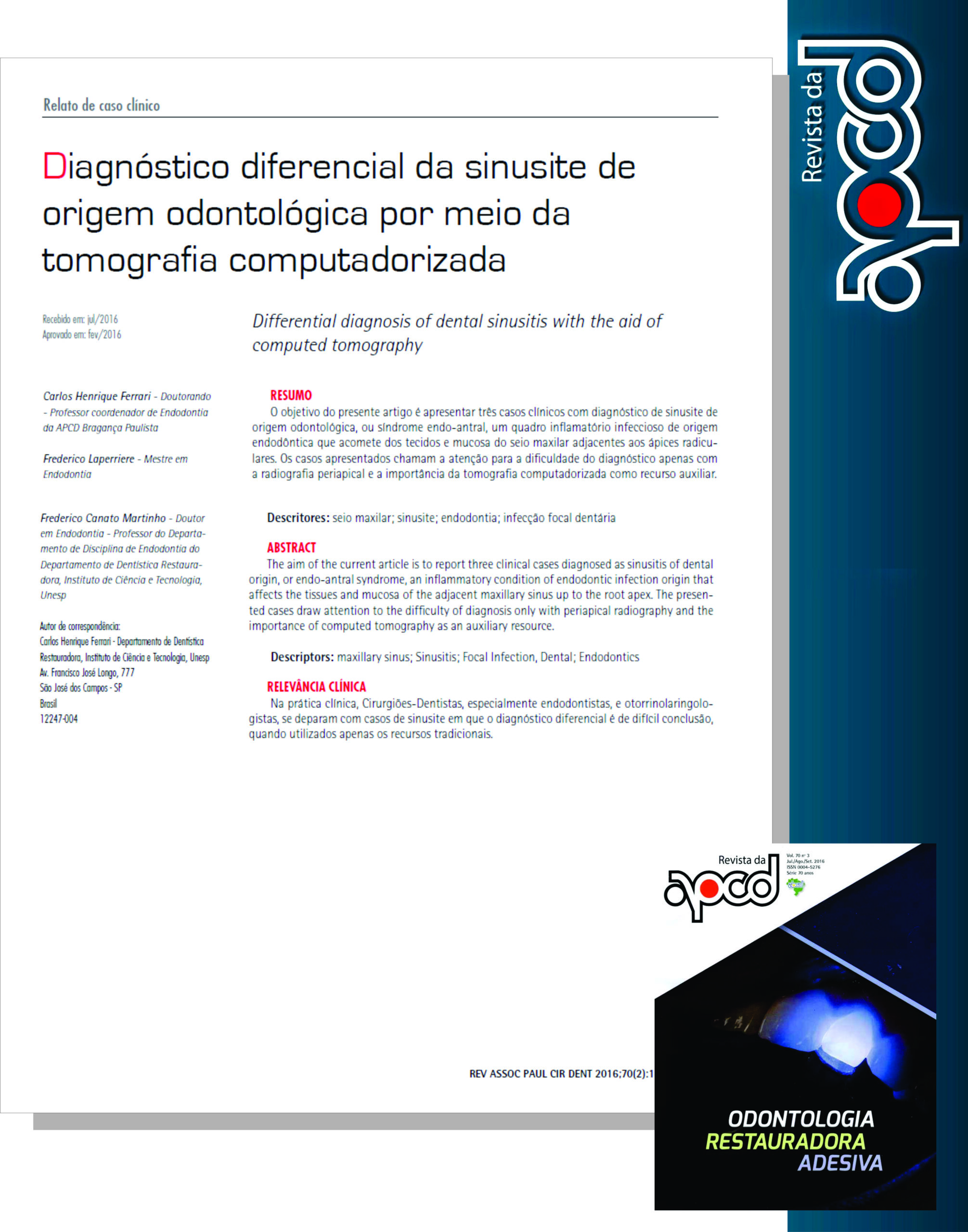
Diagnóstico diferencial da sinusite de origem odontológica por meio da tomografia computadorizada.Trabalho publicado na Revista da APCD, com o Prof. Frederico Laperriere (BH) e O Prof. Dr. Frederico Martino (Univ. de Maryland EUA)
INTRODUÇÃO
O envolvimento do seio maxilar com patologias endodônticas é descrito há muito por endodontistas e otorrinolaringologistas. A infecção proveniente do canal radicular é largamente reconhecida como causadora de inflamação das mucosas sinusais após a destruição das suas corticais ósseas. Essa capacidade de causar lesão é tão maior quanto for a proximidade dos ápices com a cortical inferior do seio. Esta distância varia muito de paciente para paciente e até mesmo em lados distintos num mesmo indivíduo. Em diversos casos, o ápice radicular se projeta no interior do seio maxilar com um mínimo de cobertura óssea, ou até mesmo nenhuma, recoberto apenas pela membrana do seio.
A origem odontogênica das sinusites é relatada em cerca de 10 a 12% dos casos. Por outro lado, a sinusite sem causa endodôntica é frequentemente reportada por pacientes e até confundida por cirurgiões dentistas, como odontológica, ressaltando a importância da correta interpretação dos achados clínicos e exames por parte dos profissionais de saúde envolvidos.
O conjunto de sinais e sintomas que definem o quadro de sinusite de origem endodôntica, é denominado síndrome endo-antral e é caracterizado por: infecção endodôntica em um dente próximo ao seio maxilar, imagem radiolúcida periapical relacionada ao dente afetado, perda da lâmina dura em volta do ápice correspondente à borda inferior radiográfica do seio maxilar, imagem radiopaca no seio maxilar sobre o ápice do dente envolvido e variação de graus de radiopacidade na imagem do seio maxilar acometido pela inflamação, comparado ao seio maxilar do lado oposto.
CASO CLÍNICO 1
Paciente sexo masculino, 56 anos, apresentou-se ao cirurgião dentista relatando dor à mastigação. Na anamnese, o paciente relatou quadros recorrentes de sinusite, sem causa aparente. Relatou também ter passado por tratamento endodôntico no dente 16 há 5 anos. O exame clínico revelou uma pequena área edemaciada na gengiva, na região da raiz mésio-vestibular do dente 16, além de dor ao teste de percussão. O exame radiográfico periapical revelou tratamento endodôntico satisfatório realizados nos dentes 15 e 16. Realizada a tomografia, observou-se no dente 16, imagem de um outro canal mésio-vestibular não tratado, rarefação óssea periapical circunscrita na raiz mésio-vestibular, rompimento da cortical inferior do seio maxilar contígua à referida raiz e velamento parcial da imagem do seio maxilar. Ressalte-se que nenhum desses achados foram observados no exame radiográfico periapical.
CASO CLÍNICO 2
Paciente sexo feminino, 40 anos, compareceu ao serviço odontológico encaminhado por outro colega devido à carie extensa no dente 17. Na anamnese não relatou qualquer sintoma relacionado à patologia endodôntica ou sinusal. No exame clínico foi constatado presença de cárie extensa na região mesial e resposta negativa aos testes de vitalidade pulpar. No exame radiográfico periapical observou-se imagem radiolúcida extensa na coroa e rarefação óssea periapical difusa aparentemente sobre a raiz palatina. O exame tomográfico revelou perda óssea e de lâmina dura contíguas à região periapical das três raízes e extenso rompimento e afastamento da cortical inferior do seio maxilar, além de imagem radiopaca compatível com inflamação da mucosa sinusal ao redor de toda área atingida.
CASO CLINICO 3
Paciente sexo masculino, 41 anos, compareceu ao consultório com relatando dor ao toque no dente 26. Na anamnese relatou que o dente havia sido tratado endodonticamente a mais de 10 anos. No exame clínico constatou-se dor à percussão. No exame radiográfico verificou-se tratamento endodôntico insatisfatório e presença de imagem radiolúcida na raiz MV. O exame tomográfico demonstrou pequena imagem radiolúcida difusão na região do ápice do canal palatino e extensa imagem radiolúcida circunscrita por cortical óssea envolvendo as raízes vestibulares, com espessamento das mucosas do seio maxilar na região contígua à lesão, além de um canal MV2 que não havia sido tratado.
https://ferrariendodontia.com.br/perfuracao-radicular-cirurgia/


