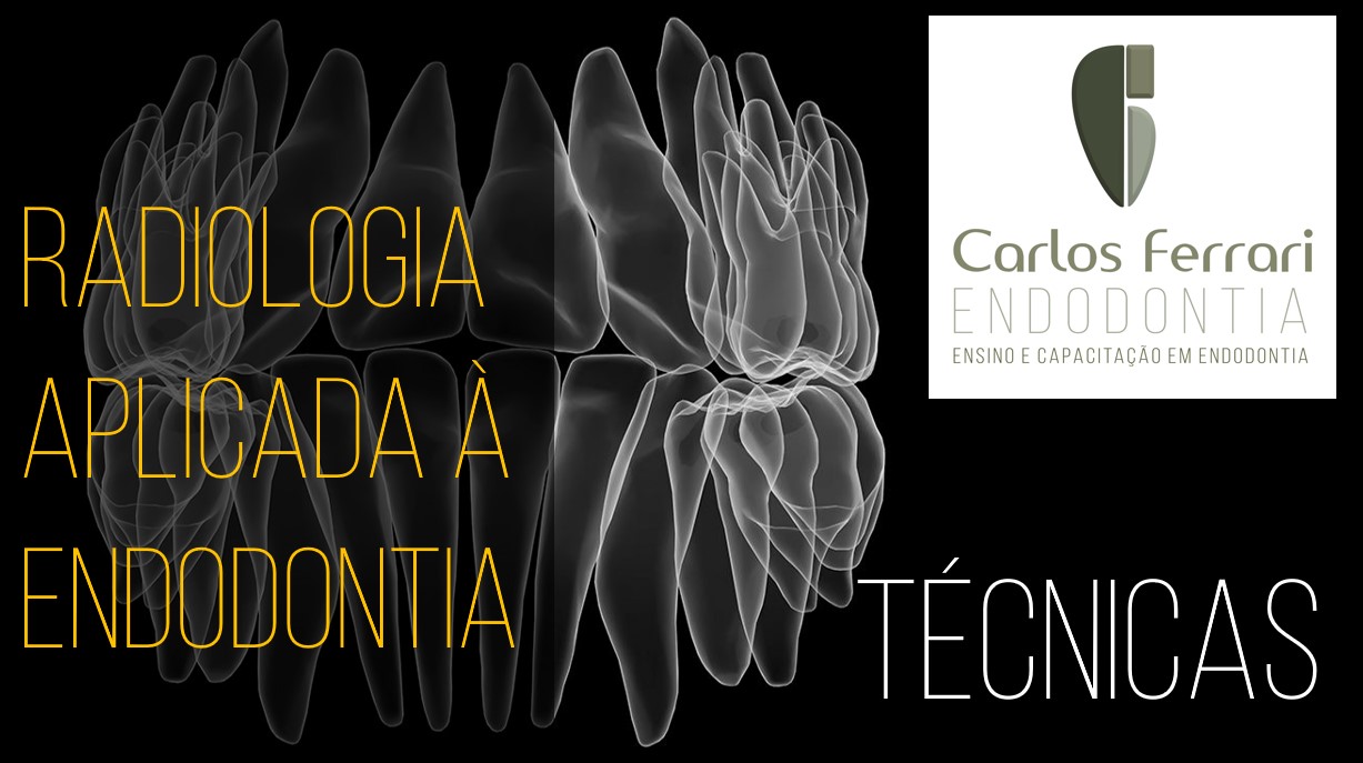
Essa segunda aula mostra a aplicação da técnica radiográfica na endodontia, com especial ênfase na técnica de Clark.
A técnica periapical é a técnica de escolha como auxiliar no diagnóstico, planejamento e proservação e a única técnica utilizada durante o tratamento endodôntico nas manobras de odontometria, prova do cone e da obturação e na avaliação final do tratamento. Por meio dela, podemos observar em uma única radiografia, coroa, a raiz e o osso periapical, bem como todas as alterações que afetam essas estruturas.
Técnicas da bissetriz e paralelismo
A técnica radiográfica na endodontia pode variar da bissetriz ao paralelismo. A técnica da bissetriz é a mais utilizada na odontologia. Tem esse nome devido ao fato de que o feixe de raios-x incide perpendicularmente à bissetriz entre o longo eixo do dente e o longo eixo do filme ou sensor que é mantido em posição no palato ou no rebordo lingual alveolar por meio do dedo do paciente em sua técnica original. Para o posicionamento do filme ou sensor, podem ser utilizados também posicionadores. radiografias em que o feixe de raios-x seja posicionado perpendicularmente ao filme ou sensor, que por sua vez está paralelo ao longo eixo do dente. Isso é possível com o uso de posicionadores que mantem o filme ou sensor paralelo ao dente e uniformizam a posição do feixe por meio de um anel guia. A técnica do paralelismo possui algumas vantagens em relação à técnica da bissetriz, como simplicidade para execução sem necessidade do posicionamento da cabeça do paciente, maior fidelidade na imagem do formato e comprimento das raízes, melhor determinação das dimensões de imagens radiolúcidas periapicais, padronização do exame para comparações posteriores na proservação, menores sobreposições de estruturas anatômicas sobre o ápice radicular e menor exposição do paciente à radiação secundária. Além disso com o uso de posicionadores é mais fácil a mudança da angulação horizontal, recurso muito utilizado na endodontia.
Técnicas periapicais em pacientes com isolamento absoluto.
A obtenção de radiografias periapicais auxiliares ao tratamento endodôntico tem como fator de complicação a presença do isolamento absoluto, o que de maneira alguma impede de que a técnica seja realizada, desde que observados certos princípios e se necessário realizadas adaptações. O primeiro aspecto a ser observado, diz respeito à tomada dos exames sem retirada do conjunto grampo, arco e lençol. A retirada de arco e lençol também não é recomendada, já que, além da possibilidade de contaminação da câmara pulpar, ainda dificulta a observação do dente e grampo. Para a execução da técnica portanto, basta que se afaste o lençol em três ou 4 pontos do arco do lado e arcadas opostos ao dente tratado. Um porta agulha ou pinça hemostática podem ser utilizados para posicionamento do filme radiográfico, quando da utilização da técnica convencional. Essa técnica permite a apreensão firme do filme, posicionamento mais simples e normalmente uma melhor aceitação de pacientes com náuseas, já que fica responsável por segurar o posicionador e removê-lo, se necessário. Há também no mercado, posicionadores que permitem a realização de radiografias com isolamento absoluto na técnica do paralelismo. O bloco de mordida é localizado anteriormente ao posicionador do filme radiográfico ou sensor e seu respectivo anel de localização. Esses dispositivos permitem maior padronização dos exames, inclusive auxiliando na realização de técnicas com alteração da angulação do feixe de raios-x em relação à tomada ortorradial.
Técnica de Clark.
A técnica radiográfica na endodontia, com maior utilização é a técnica de Clark é no diagnóstico (reabsorções radiculares e perfurações), planejamento (anatomia e número de canais radiculares), observação das limas na odontometria, ajuste dos cones de guta percha, avaliação final do tratamento e proservação, por meio da realização de tomadas mesiorradiais e distorradiais, com o intuito de dissociação entre as raízes e canais que estão num mesmo plano em direção vestíbulo-lingual ou da observação da anatomia em raízes achatadas, no mesmo sentido. A maior utilização, portanto, se dá nas seguintes raízes: prés-molares superiores, raiz mésio-vestibular dos molares superiores, anteriores inferiores e raiz mesial dos molares inferiores. Outra utilização importante é para dissociação da imagem da região apical de outras imagens que possam estar sobrepostas, como corpos estranhos ou estruturas anatômicas (canal mandibular, forame mentoniano, canal incisivo, seio maxilar).
Técnica de rastreamento de fístula.
A presença de uma fístula, comum em casos de abscesso periapical assintomático, pode causar dúvidas quanto ao dente causal. Não raramente em casos de sua presença encontram-se mais de um dente com diagnóstico de necrose pulpar e presença de rarefação periapical, porém é importante o correto diagnóstico sobre sua origem.