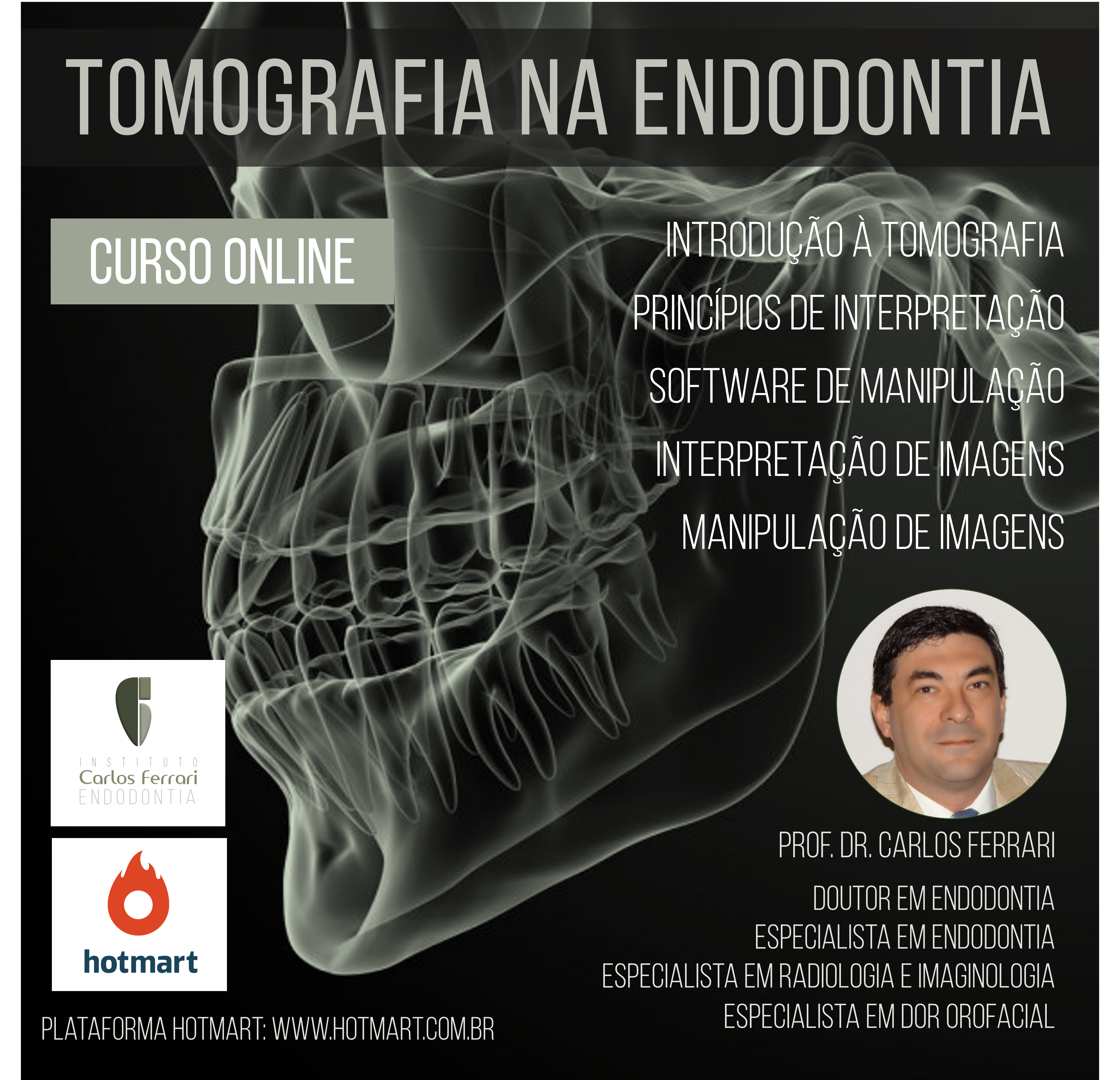
Tomografia na endodontia, curso online na plataforma Hotmart, está disponível, com 8 horas aula de duração.
A tomografia cone beam, hoje é realidade para o planejamento, tratamento e proservação dos casos clínicos na rotina endodôntica. Cada vez mais os profissionais buscam nesse recurso um apoio ao trabalho e ao melhor diagnóstico e tratamento. Portanto é fundamental a capacitação para esse recurso, que vai muito além de receber as imagens impressas na pasta de documentação.
Aplicações da tomografia na endodontia
A tomografia na endodontia tem aplicações nas diversas fases do tratamento, do diagnóstico até a proservação dos casos. Um exame tomográfico é capaz de fornecer mais informações do que a radiografia periapical e é capaz de fazer com que de 30 a 60% dos casos, haja mudança de diagnóstico ou conduta do profissional em relação àquela adotada somente com o primeiro exame.
A tomografia é útil para a observação tridimensional da morfologia dental, revelando com muito mais fidelidade a câmara pulpar, com assoalho e entrada dos canais e o número e formato dos canais radiculares e útil na detecção de canais adicionais. Além disso, dentes com anatomia anormal, podem ser muito melhor entendidos com o auxílio do exame, além da observação anomalias e malformações. Em retratamentos, é de suma importância na detecção de canais não tratados, situação comum em raízes achatadas em sentido mesiodistal, citadas anteriormente.
A tomografia na endodontia também pode ser utilizada para detecção de trincas ou fraturas radiculares.
Em caso de reabsorções radiculares, a tomografia é um meio mais eficaz que as radiografias intra orais para localização e dimensionamento das lacuna. Além disso, somente um exame tridimensional pode avaliar as comunicações das reabsorções com o canal radicular, em casos de reabsorção externa, ou com os tecidos periodontais, em caso de reabsorção interna.
Outra aplicação da tomografia na endodontia, com maior eficácia na detecção das imagens hipodensas em relação ao exame radiográfico diz respeito à observação das lesões periapicais, tanto no diagnóstico, como no diagnóstico diferencial entre lesões não endodônticas, no planejamento cirúrgico e na proservação. A tomografia revela imagens no periápice mais precocemente, em estágios ainda não observáveis na radiografia periapical e com maior percentual de detecção.
Quando as imagens hipodensas são localizadas em dentes superiores posteriores, o exame pode ser fundamental no diagnóstico diferencial da sinusite de origem odontogênica, ao revelar a comunicação das lesões com o seio maxilar, além de rompimento de corticais e espessamento das mucosas adjacente. Essa condição é comum e pouco diagnosticada tanto por cirurgiões dentistas, quanto por médicos.
Uma utilização da tomografia , útil tanto para o planejamento de cirurgia paraendodôntica como na prevenção de acidentes em tratamentos endodônticos invasivos além do forame apical, em que se realizam manobras de patência e alargamento foraminal, é a avaliação das distâncias entre ápices e estruturas anatômicas importantes, como seio maxilar e canal mandibular, bem como a observação prévia ao tratamento de fenestrações apicais. Portanto, a observação dessa última também pode ser útil para o diagnóstico de dores pós operatórias de longa duração, sem causa aparente
Em traumatismos dentais, a tomografia é útil para revelar fraturas de coroa e/ou radiculares e ósseas, bem como as imagens de espaço alveolar vazio, típicos das lesões envolvendo deslocamento dentário . A tomografia mostrou-se mais eficiente que as panorâmicas na observação de fraturas ósseas e alveolares e que as radiografias periapicais digitais na detecção de fraturas radiculares horizontais, inclusive quando a modificação de angulação horizontal dos raios-x não foi capaz de revelar a fratura.
Em casos de dentes com câmara pulpar e canal radicular atresiados por mineralização, situação comum em dentes anteriores com histórico de traumatismo, a tomografia, aliada à tecnologia de prototipagem, pode ser utilizada por meio de guias confeccionados em impressoras 3D, para nortear o acesso de uma broca cirúrgica até a região periapical. Tal tratamento é denominado acesso endodôntico guiado .
Outras aplicações em que a tomografia também se mostra útil são a localização de corpos estranhos, instrumentos fraturados, perfurações, avaliação de extravasamento de cone de guta percha e/ou cimento obturador e nos vários campos de pesquisa na endodontia.
curso de tomografia na endodontia na plataforma hotmart
O curso vai capacitar o acadêmico, cirurgião dentista ou especialista em endodontia, para conhecer, indicar e interpretar a tomografia cone beam com finalidade endodôntica, e é composto de módulos teórico e prático, em que o aluno poderá manipular imagens no software.